Class II Deep Margin Elevation Following Single-Visit Endodontic Treatment Under Strict Isolation
Adhesive-Driven Rehabilitation of a Mandibular First Molar
Author: Dr. Hamza Zahid
Microscopic Restorative Dentist
Abstract
Deep proximal caries extending subgingivally present both biological and restorative challenges. When combined with pulpal involvement, management requires a seamless transition from endodontics to adhesive rehabilitation. This article presents a single-visit endodontic treatment of a mandibular first molar followed by Class II Deep Margin Elevation (DME) and definitive composite restoration under strict rubber dam isolation. The case demonstrates how adhesive principles, matrix control, and margin relocation can preserve periodontal health while ensuring structural integrity and long-term predictability.
Introduction
Restoring teeth with deep proximal margins has historically involved surgical crown lengthening or orthodontic extrusion. However, Deep Margin Elevation (DME), first described by Dietschi and Spreafico, allows relocation of subgingival margins coronally using bonded composite, facilitating adhesive restoration while maintaining periodontal stability.
When performed under strict isolation and correct adhesive protocols, DME provides:
- Improved visibility and accessibility
- Predictable bonding
- Controlled proximal contours
- Respect for biologic width
This case integrates single-visit endodontics with adhesive-driven DME to restore function and anatomy conservatively.
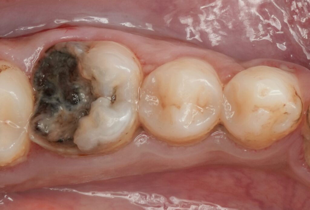
Diagnosis and Treatment Planning
Clinical findings:
- Extensive distal caries on mandibular first molar
- Subgingival proximal margin
- Pulpal involvement
- Tenderness on percussion
- Radiographic evidence of deep carious lesion approaching pulp
Treatment Plan:
- Single-visit root canal therapy
- Immediate adhesive coronal seal
- Class II Deep Margin Elevation
- Direct composite restoration
Rationale: Immediate adhesive sealing reduces coronal leakage and improves long-term prognosis of endodontically treated teeth.
Step-by-Step Clinical Protocol
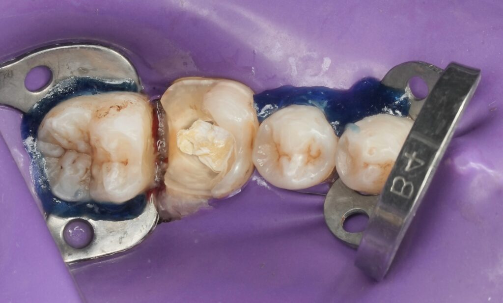
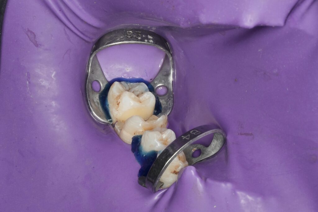
1. Rubber Dam Isolation
Strict isolation was achieved using clamp stabilization and liquid dam reinforcement.
Why this matters:
Adhesive dentistry is moisture-sensitive. Isolation is non-negotiable for predictable bonding (Mounce, 2004).
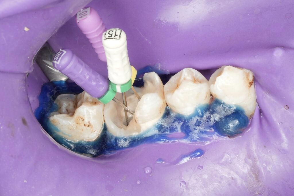
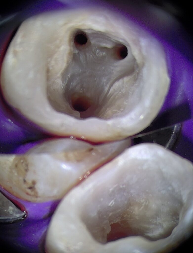
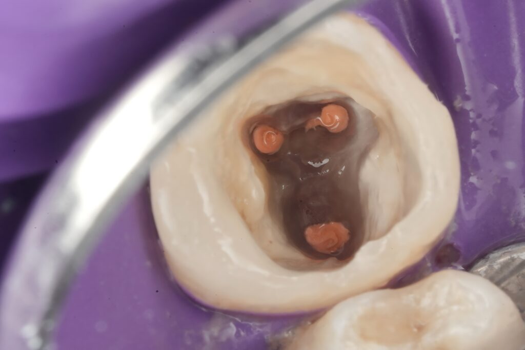
2. Access Opening and Canal Identification
- Conservative access cavity preparation
- Identification of all canals
- Working length determination using apex locator and radiographic confirmation
3. Cleaning and Shaping
- Rotary NiTi instrumentation
- Copious irrigation with sodium hypochlorite
- Final rinse protocol
Proper debridement ensures microbial control, which is the primary determinant of endodontic success (Sjögren et al., 1997).
4. Obturation
- Warm vertical compaction technique
- Dense 3D obturation
- Radiographic confirmation
Immediate coronal sealing followed.
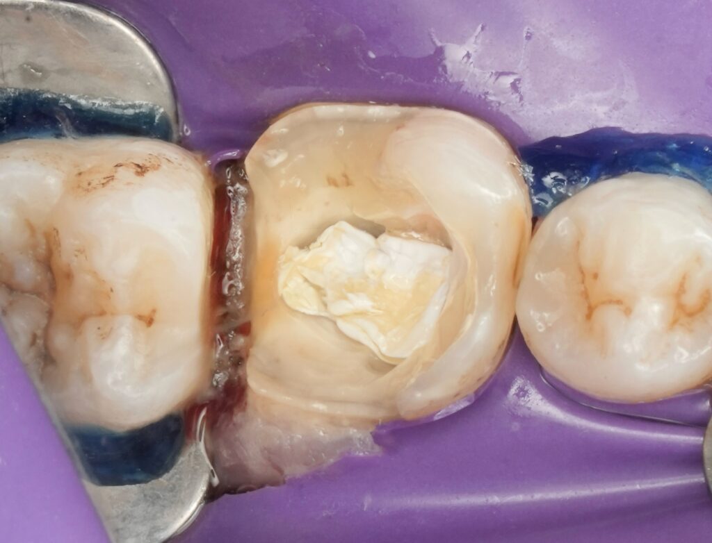
5. Immediate Coronal Seal
A bonded composite core was placed to prevent microleakage. Literature consistently supports immediate coronal sealing to improve survival rates (Ray & Trope, 1995).
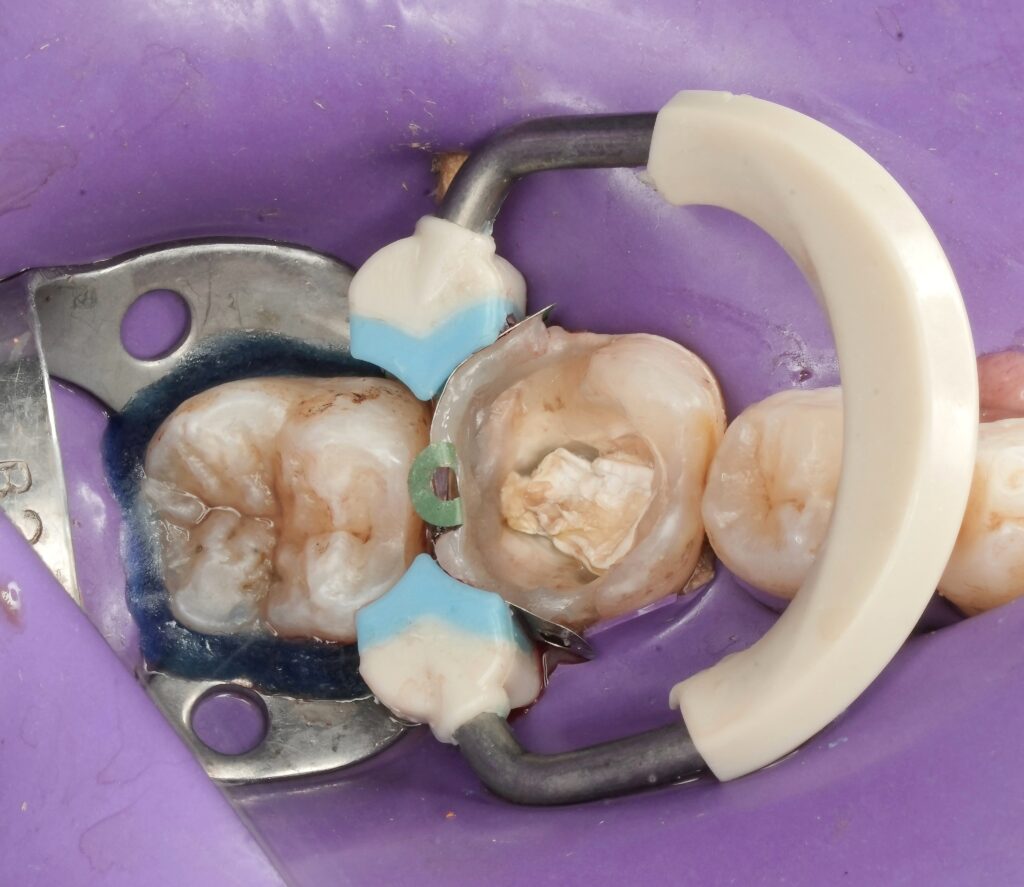
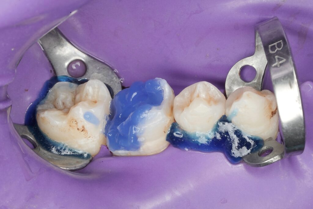
6. Proximal Matrix Placement
A sectional matrix system was used with wedge adaptation to:
- Achieve tight contact
- Protect gingival tissues
- Control marginal emergence
Matrix precision is critical for periodontal stability.
7. Deep Margin Elevation (DME)
The deep distal margin was elevated coronally using incremental composite placement.
Protocol:
- Selective enamel etching
- Adhesive application
- Flowable composite base
- Layered composite build-up
The margin was relocated supragingivally, allowing ideal finishing and polishing.
DME allows preservation of tooth structure while avoiding surgical intervention (Dietschi & Spreafico, 1998).
8. Occlusal Anatomy Reconstruction
Using layered composite technique:
- Dentin layer for opacity
- Enamel layer for translucency
- Cusp-by-cusp buildup
- Precise occlusal morphology reproduction
Anatomical reproduction improves stress distribution and functional longevity.
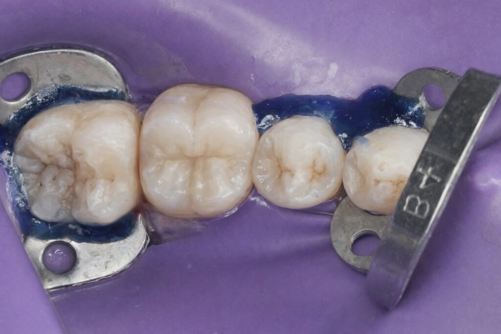
9. Finishing and Polishing
- Fine diamond refinement
- Multi-step polishing system
- Verification of proximal contact
- Occlusal adjustment
Proper finishing reduces plaque retention and enhances marginal integrity.
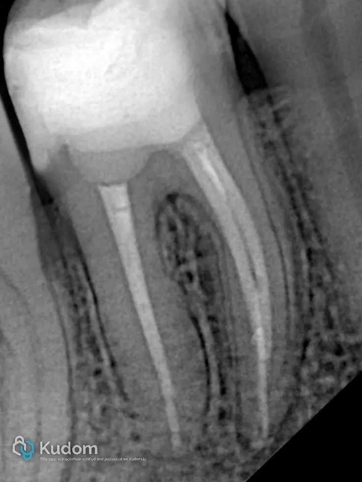
Radiographic Outcome
Postoperative radiographs confirmed:
- Dense obturation
- Well-adapted coronal seal
- Proper proximal contour
- No marginal overhang
Biological Considerations
Respecting the supracrestal tissue attachment (formerly biologic width) is essential.
DME is acceptable when:
- Isolation is achievable
- Margins do not invade attachment apparatus
- Periodontal inflammation is controlled
Long-term studies show stable periodontal outcomes when margins are well-contoured and polished (Ferrari et al., 2018).
Discussion
This case highlights key restorative principles:
- Endodontics and restorative dentistry must be integrated, not separated.
- Immediate adhesive sealing improves survival.
- Deep margins can be predictably relocated without surgical trauma.
- Rubber dam isolation remains the cornerstone of success.
For endodontically treated molars, structural preservation combined with adhesive reinforcement offers a minimally invasive alternative to full-coverage crowns in selected cases.
Conclusion
From deep subgingival caries to complete adhesive rehabilitation, this case demonstrates that precision, isolation, and respect for biology allow predictable outcomes.
Class II DME under strict isolation transforms a compromised tooth into a functionally stable and aesthetically integrated restoration — without unnecessary surgical intervention.
Modern restorative dentistry is not about replacing structure.
It is about preserving what can be preserved — and rebuilding what must be rebuilt — intelligently.
References
- Dietschi D, Spreafico R. Current clinical concepts for adhesive cementation of tooth-colored posterior restorations. Pract Periodontics Aesthet Dent. 1998.
- Ray HA, Trope M. Periapical status of endodontically treated teeth in relation to the quality of coronal restoration. Int Endod J. 1995.
- Sjögren U et al. Influence of infection at the time of root filling on the outcome of endodontic treatment. Int Endod J. 1997.
- Mounce R. Rubber dam isolation in endodontics. Dent Today. 2004.
- Ferrari M et al. Deep margin elevation: clinical evaluation and periodontal considerations. J Adhes Dent. 2018.
- Magne P, Spreafico R. Deep margin elevation: A paradigm shift. Int J Esthet Dent.
- Bresser RA et al. Adhesive restoration of endodontically treated teeth: survival analysis. J Dent Res.
{"tiktok_developers_3p_anchor_params":"{"template_id":"","client_key":"awgvo7gzpeas2ho6","filter_id":[],"capability_extra_v2":{"edit":[{"panel":"hd_quality_picture"}]},"capability_key":["edit"]}","data":{"product":"hypic","activityName":"","filterId":"","imageEffectId":"","enter_from":"enter_launch","stickerId":"","playId":"","pictureId":"59218B2B-126D-4D88-8A01-AA7AADB6CBAF","infoStickerId":"","appversion":"8.1.0","os":"ios"},"source_type":"hypic"}
Share on: