Subtitle
Preserving pulp vitality through controlled caries removal, strict isolation, and adhesive reconstruction
Introduction
Management of deep carious lesions in posterior teeth has shifted toward minimally invasive strategies focused on pulp preservation. Direct pulp capping, when performed under strict isolation with proper case selection, can maintain vitality and avoid endodontic intervention.
This case demonstrates a biomimetic approach combining vital pulp therapy with adhesive Class II restoration, emphasizing peripheral seal zone integrity and controlled layering.
Diagnosis & Treatment Planning
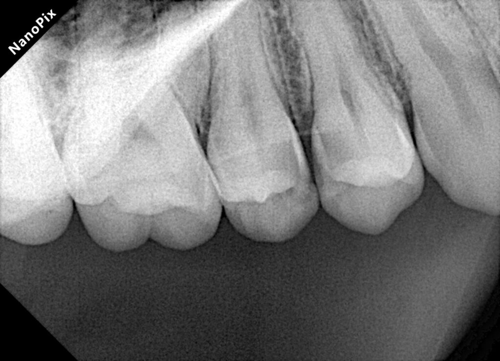
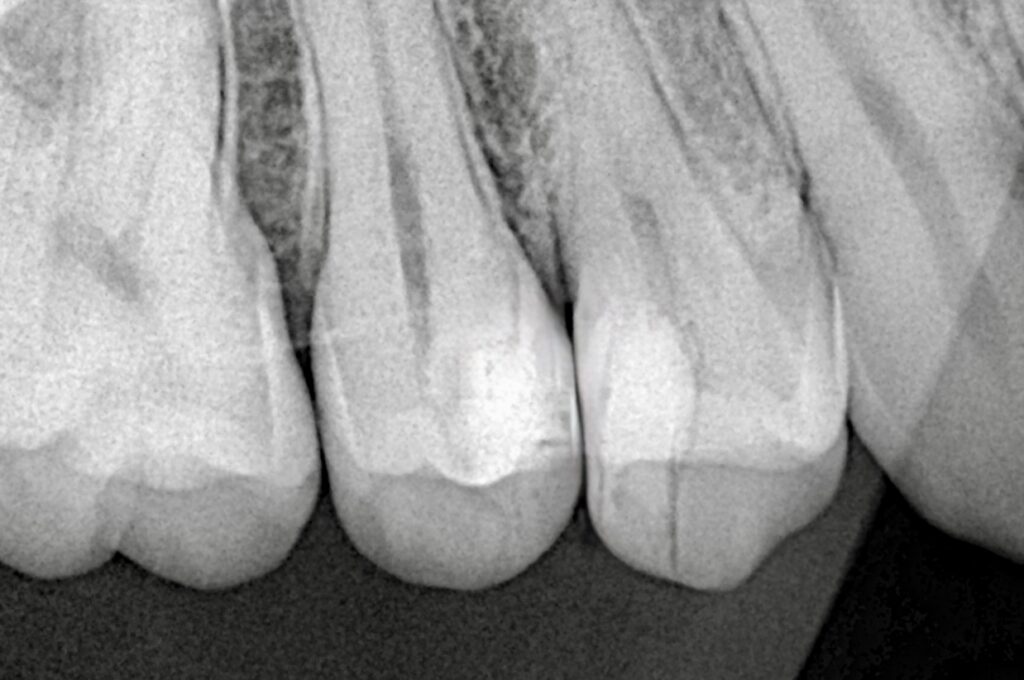
- Deep proximal caries in premolar
- Vital tooth with no signs of irreversible pulpitis
- Radiographic evaluation suggestive of deep dentinal involvement approaching pulp
Plan:
- Selective caries removal
- Direct pulp capping
- Immediate adhesive restoration with cuspal support consideration
Clinical Procedure
Step 1 – Isolation
Rubber dam isolation achieved to ensure a clean, moisture-free field. Proper inversion and retraction were critical due to proximal extension.
Step 2 – Caries Removal
- Peripheral seal zone completely cleaned
- Selective caries removal near pulp to avoid unnecessary exposure
- Controlled exposure occurred due to deep lesion
Step 3 – Hemostasis & Disinfection
- Hemostasis achieved within a few minutes
- Gentle irrigation (NaOCl/Saline) used for disinfection
- A stable clot-free environment confirmed before proceeding
Step 4 – Direct Pulp Capping
- MTA placed over exposure site
- Ensured proper sealing and thickness
- Allowed initial setting before restoration
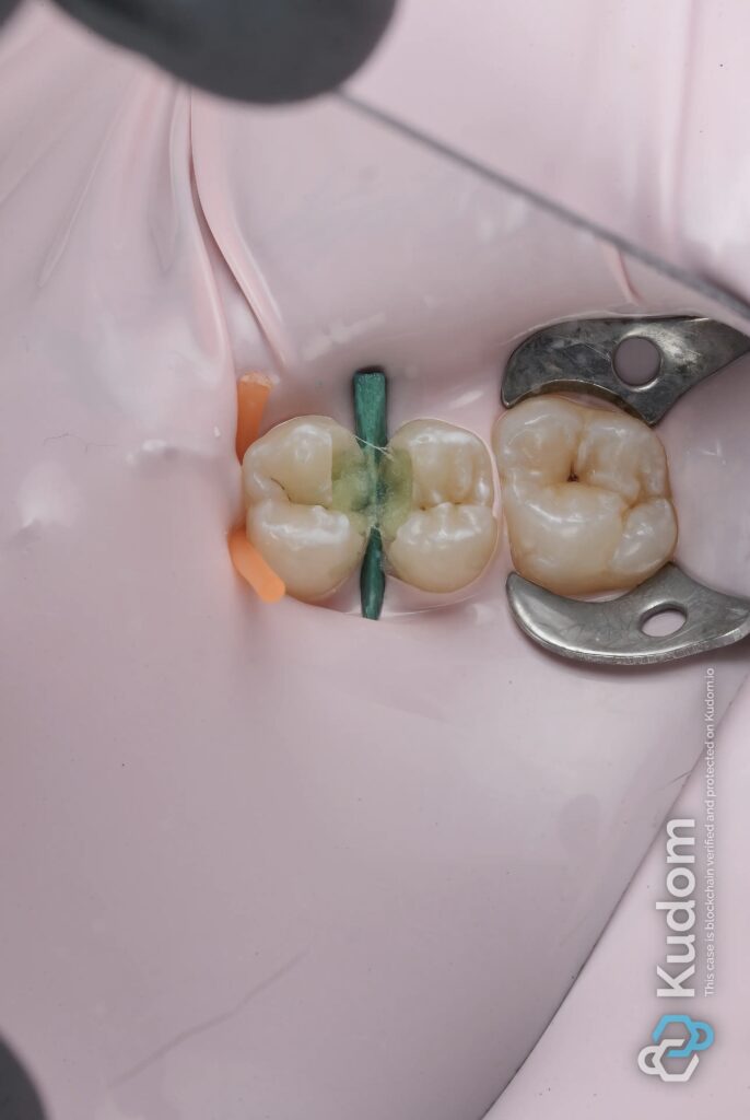
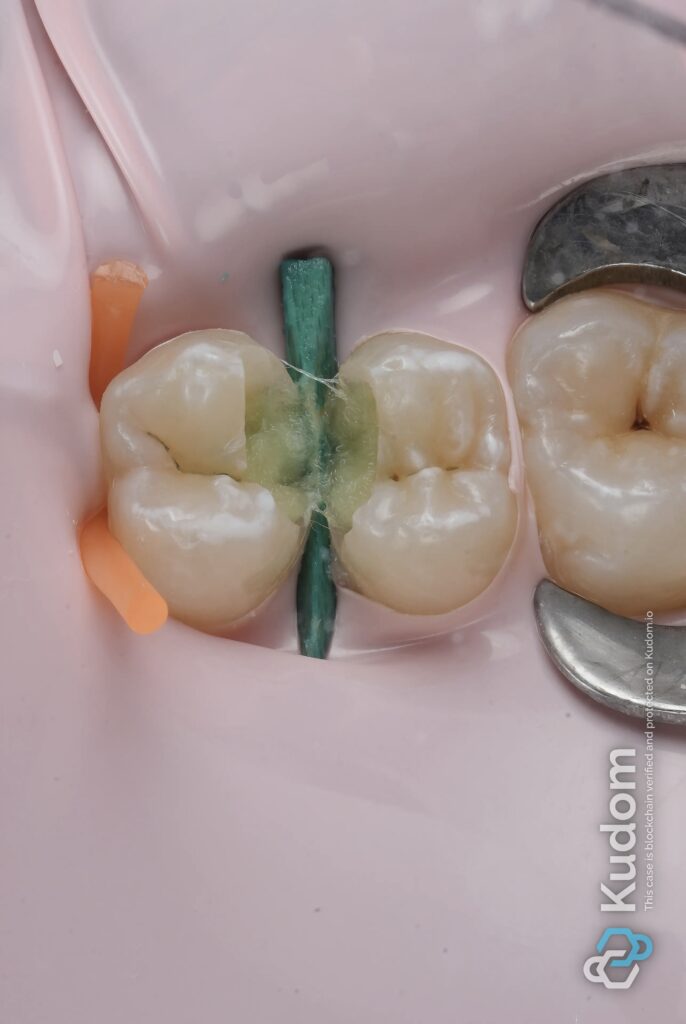
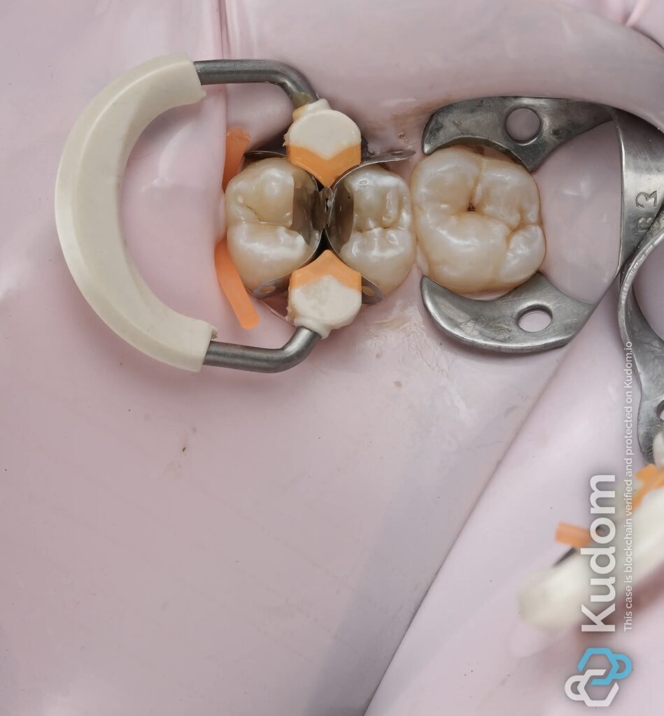
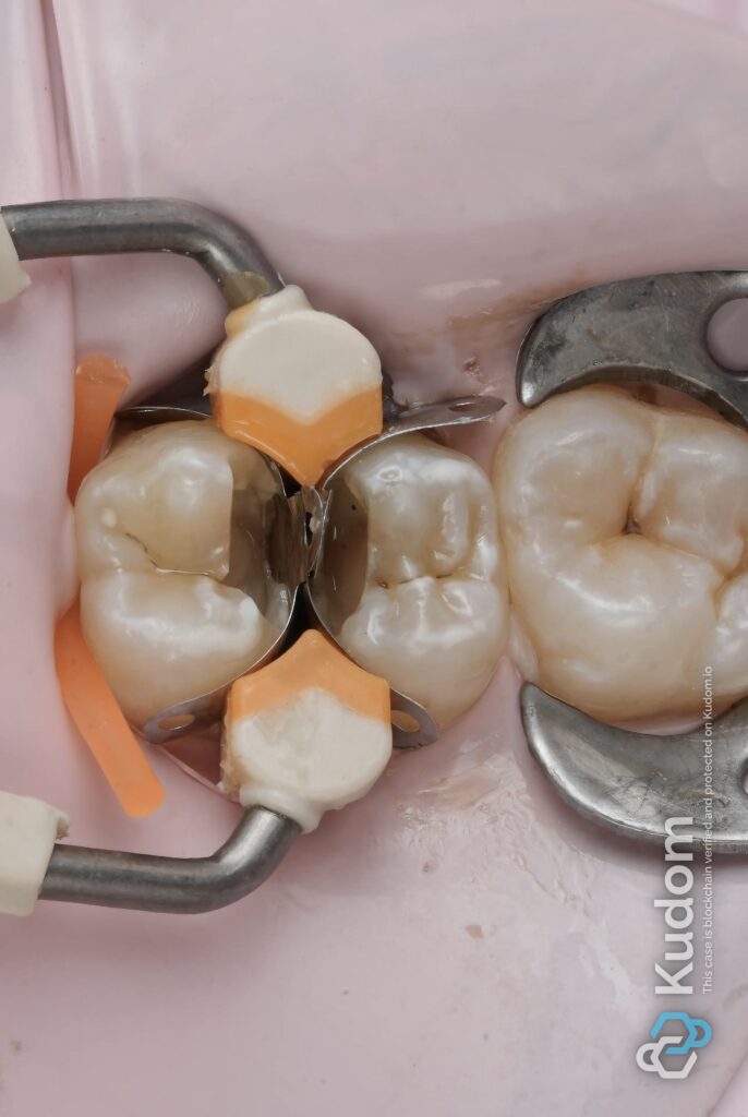
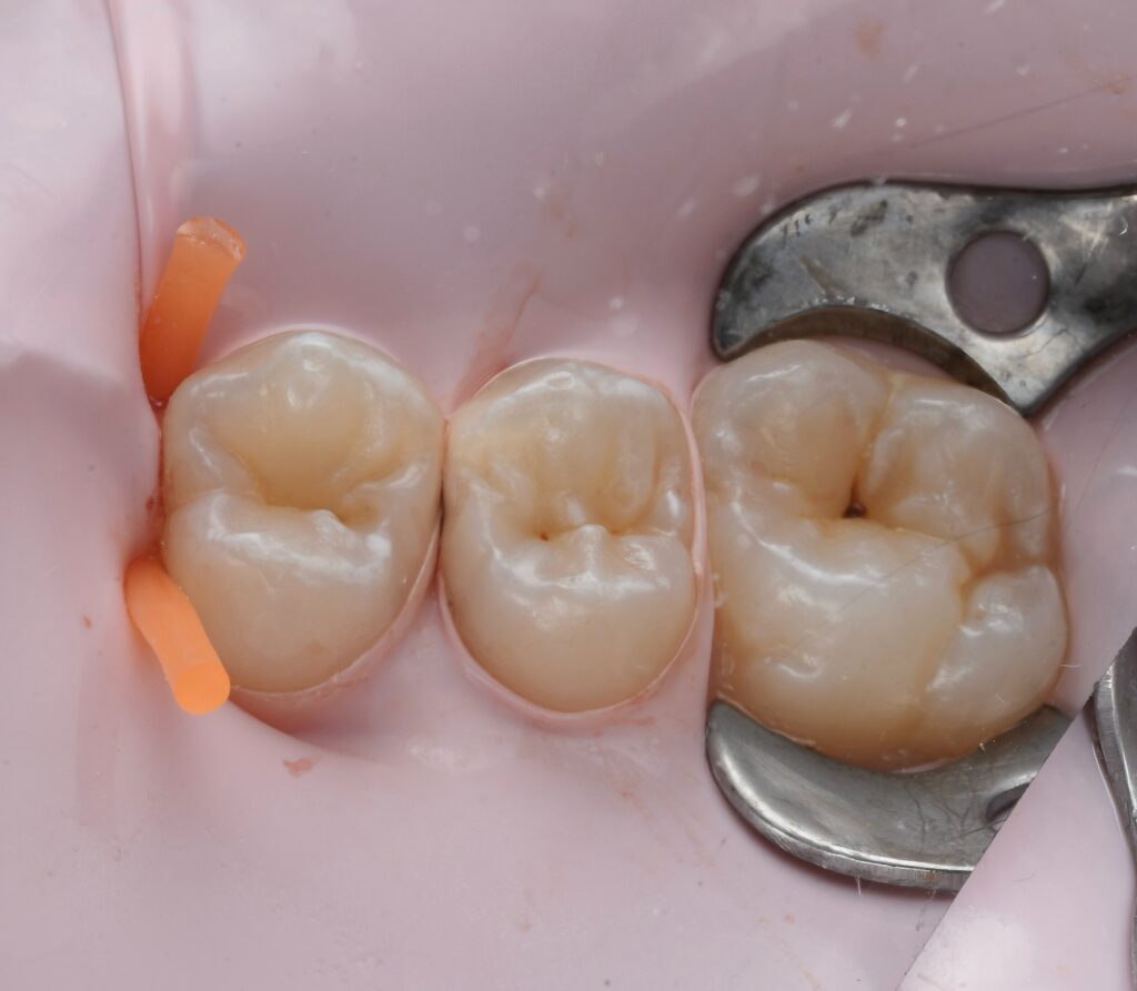
Step 5 – Matrix Placement
- Sectional matrix system with proper wedging
- Achieved tight proximal seal and contour
- Critical for contact and marginal adaptation
Step 6 – Adhesive Protocol
- Selective etch technique
- Application of bonding system (e.g., Clearfil SE Bond 2)
- Proper air thinning and curing
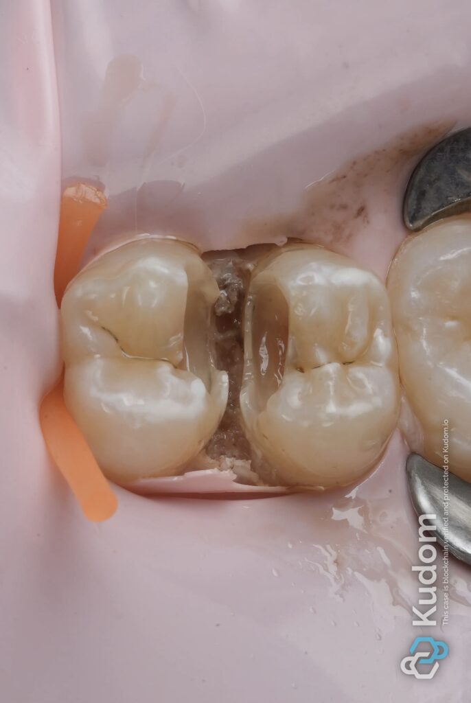
Step 7 – Composite Build-Up
- Incremental layering technique
- Emphasis on:
- Proximal wall first
- Cusp-by-cusp reconstruction
- Anatomical occlusal morphology
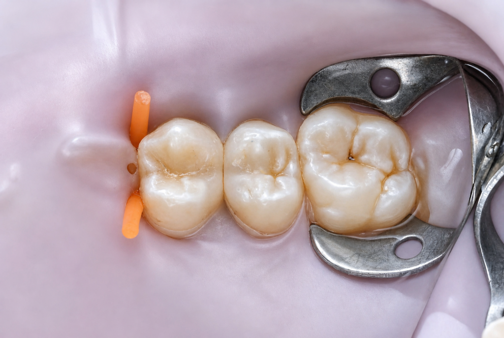
Step 8 – Finishing & Polishing
- Occlusal refinement
- Polishing to achieve smooth margins and natural luster
Key Clinical Points
- Peripheral seal zone is non-negotiable for long-term success
- Rubber dam isolation directly influences prognosis
- Controlled pulp exposure with proper capping material can maintain vitality
- Matrix adaptation defines success in Class II restorations
- Biomimetic layering restores strength and function
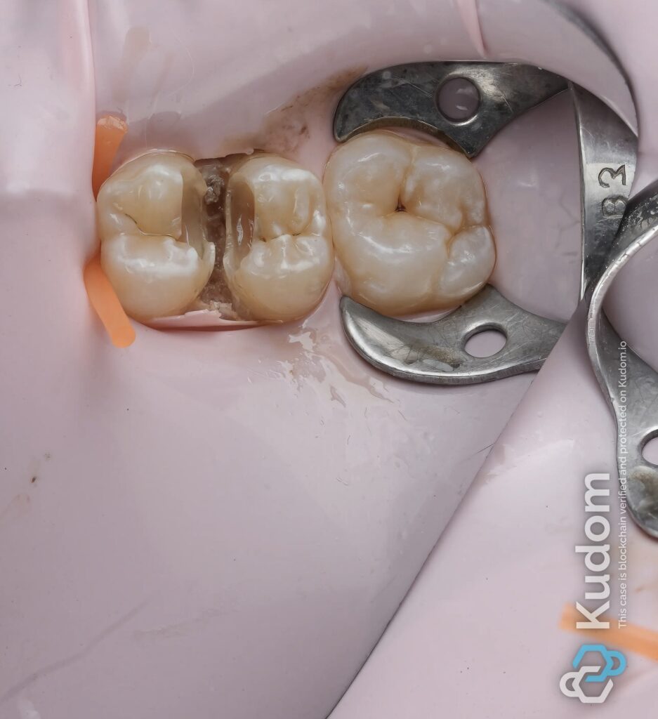
Outcome
- Functional and aesthetic restoration achieved
- Proper proximal contact and occlusal anatomy
- Tooth preserved without need for endodontic treatment
Discussion
Direct pulp capping remains a predictable option when proper protocols are followed. The success of such cases depends heavily on isolation, disinfection, and sealing ability of the restorative material.
Biomimetic dentistry plays a key role by restoring structural integrity while preserving remaining tooth structure. The combination of MTA and adhesive composite allows both biological healing and mechanical reinforcement.
🔹
Conclusion
Preserving pulp vitality should always be prioritized when clinically feasible. With correct technique and material selection, even deep lesions can be managed conservatively with long-term success.
🔹
References (for Zerodonto credibility)
- Bogen G et al. (2008). Direct pulp capping with MTA
- Hilton TJ (2009). Keys to successful pulp capping
- Murray PE et al. (2002). Pulp healing and regeneration
- Magne P, Spreafico R. Biomimetic Restorative Dentistry
- Schwendicke F et al. Selective caries removal concepts
{"tiktok_developers_3p_anchor_params":"{"template_id":"","capability_extra_v2":{"edit":[{"panel":"hd_quality_picture"}]},"capability_key":["edit"],"filter_id":[],"client_key":"awgvo7gzpeas2ho6"}","data":{"infoStickerId":"","pictureId":"0E266C06-2570-4CD1-A8DD-23F32936FBD8","playId":"","appversion":"8.6.0","imageEffectId":"","filterId":"","activityName":"","enter_from":"enter_launch","os":"ios","product":"hypic","stickerId":""},"source_type":"hypic"}
Share on: