An implant-supported restoration in the esthetic zone, and anywhere else for that matter, must replicate a natural healthy tooth not only from a functional point of view, but also from an esthetic point of view. Pink esthetic is a vital factor for the overall esthetic success of dental implant restorations. To obtain an esthetic and predictable gingival architecture around implant-supported crowns, interdental papilla plays an important role.
The absence of the interplant papilla leads to cosmetic deformities, phonetic difficulty and food impaction. Reconstructing a predictable peri-implant papilla is one of the most complex and challenging aspects of implant dentistry, let alone inter-implant papilla.
Case Report:
The purpose of this report is to show 18 months follow up of a V-shaped tuberosity connective tissue graft to improve the interplant papilla between two implant-supported restorations in the upper right central incisor and lateral incisor.
Conclusion
The papilla augmentation technique presented involves careful microsurgical dissection of the papilla and the addition of dense connective tissue that can be completely surrounded by a vascular supply to enhance graft survival. Like described by the literature, this kind of grafts tends to improve with time as seen in this case.
Bibliography:
Froum S, Lagoudis M, Rojas GM, Suzuki T, Cho SC. New Surgical Protocol to Create Interimplant Papilla: The Preliminary Results of a Case Series. Int J Periodontics Restorative Dent. 2016 Mar-Apr;36(2):161-8.
Nordland WP, Sandhu HS, Perio C. Microsurgical technique for augmentation of the interdental papilla: three case reports. Int J Periodontics Restorative Dent. 2008 Dec;28(6):543-9. PMID: 19146049.
Azzi R, Etienne D, Takei H, Fenech P. Surgical thickening of the existing gingiva and reconstruction of interdental papillae around implant-supported restorations. Int J Periodontics Restorative Dent. 2002 Feb;22(1):71-7.

Initial, frontal view

New Provisionals

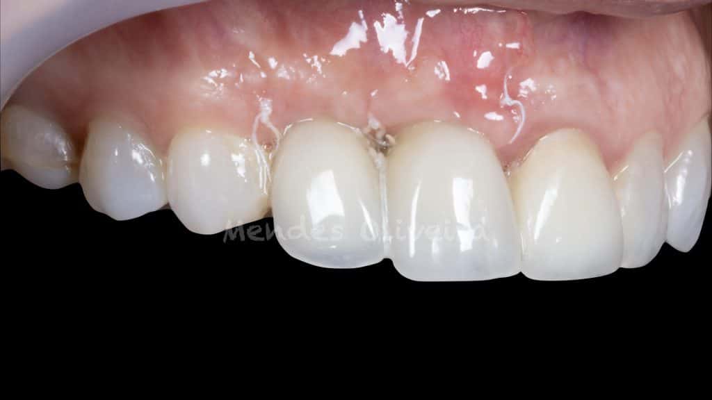
New provisionals, lateral view

Tuberosity CTG adapted to the site

Immediate result after suture

Suture removal, frontal view
Suture removal, lateral view

Frontal view after suture removal

Lateral view after suture removal
Frontal view at 2 months follow up

Lateral view at 2 months follow-up, showing interimplant space more closed

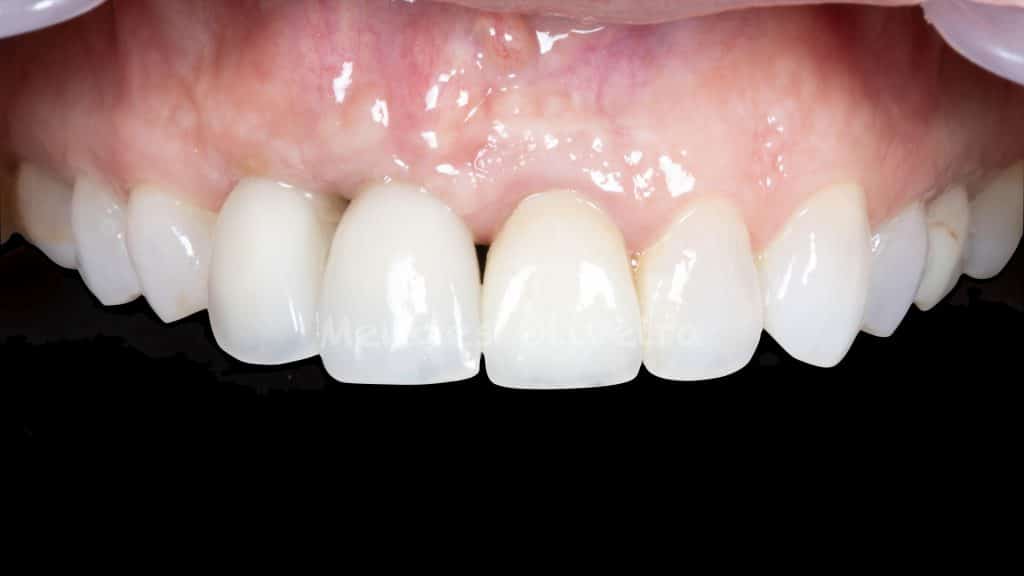
Frontal view at 18 months follow up
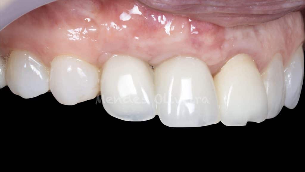
Lateral view at 18 months follow-up showing full

Before and after
Share on: