Regarding VPT :
As mentioned in the previous posts, the primary goal of VPT is to create optimal conditions for pulp tissue repair and preservation.
Following several steps, starting with the correct diagnosis to ensure the pulp still vital , the AAE current diagnostic terminology assigns a vital pulp to the one of the three categories:”normal”, “reversible pulpitis ” or “irreversible pulpitis ” (wich could be symptomatic or asymptomatic ).
In this case, the female patient was 24 years old, came to a clinic with severe pain on diagnosis :
No periapical changes on x ray detected
– No tenderness to percusion
On cold test, the tooth responed with severe pain, indicating the diagnosis of irreversible pulpitis
Isolation with rubberdam to prevent any contamination , strict aseptic procedures.
Hemostasis is achieved by using sodium hypochlorite. After Hemostasis, the pulp wound appears homogeneous, and blood filled pulp tissue
MTA placed over the pulp tissue and covered by RMGIC, and the final restoration was following the principles of adhesive dentistry
@ricucci.domenico
IDS with G2 bond
Ever x posterior for dentin replacement
Direct composite restoration

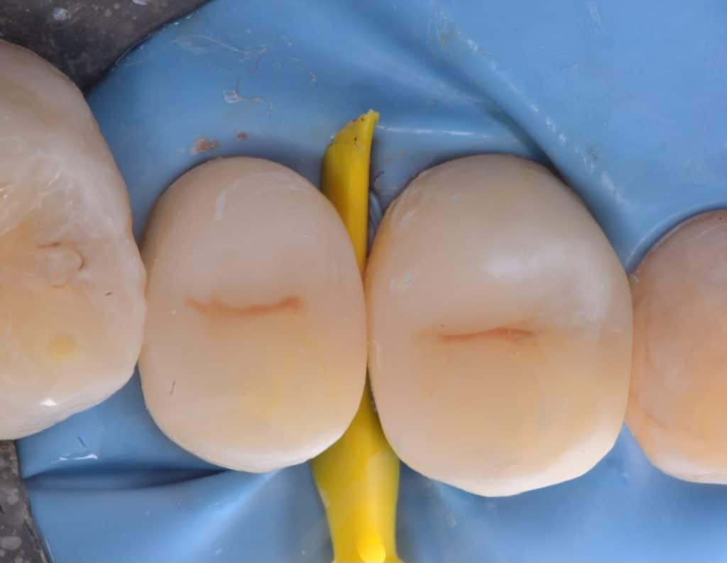
Initial situation was badly carious upper premolars with severe pain,after caries removal the pulp exposed due to the extent of carious lesion ,in attempt to saving the vitality of the teeth I decided to go with VPT as it the most recent and conservative treatment

After Hemostasis with sodium hypochlorite the pulp wound appeared continuous homogenous blood filled pulp tissue, no signs of necrosis, MTA placed over the pulp wound and direct composite done to get better coronal Sealing that increase the successful of treatment

MTA covered with light cure flowable GIC , Unsupported enamel removed, final cavity designed

Step by step , IDS with G2 bond , Ever x posterior for dentin replacement, direct composite restoration done for the second premolar , finished and polished, then first premolar followed .
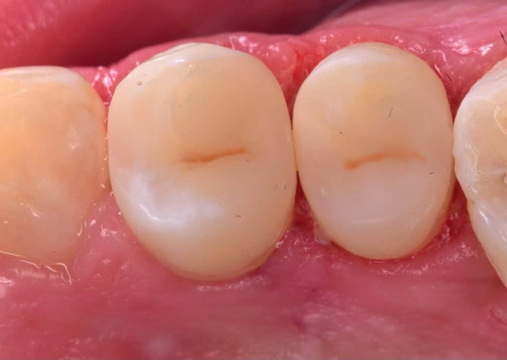
The two premolars are completed

Final result
Immediate result

Contact


Post operative x ray
Share on: