Patient, 32 years old, presented with gingival hypertrophy in the regions of teeth 12 and 11, which led to the displacement of these dental elements.
In 2013, an initial procedure was performed to reduce the hypertrophy, including a histological biopsy that returned negative results.
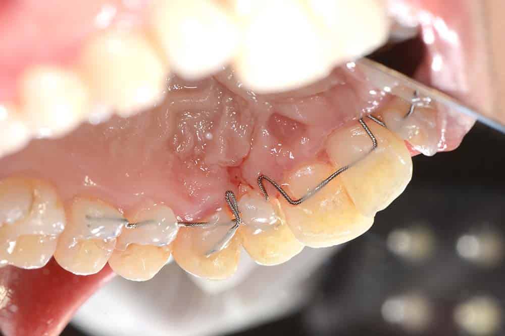
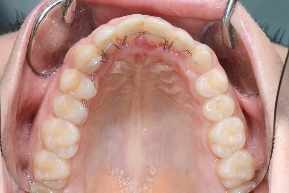
Subsequently, the patient has undegone a bracketless fixed orthodontics treatment and the tooth were aligned by means of an active retainer placed on the internal surface of the teeth.
The final outcome was satisfactory for the patient and remained stable as long as the patient maintained hygiene appointments every three months.
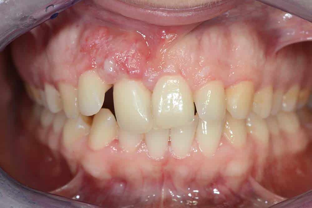
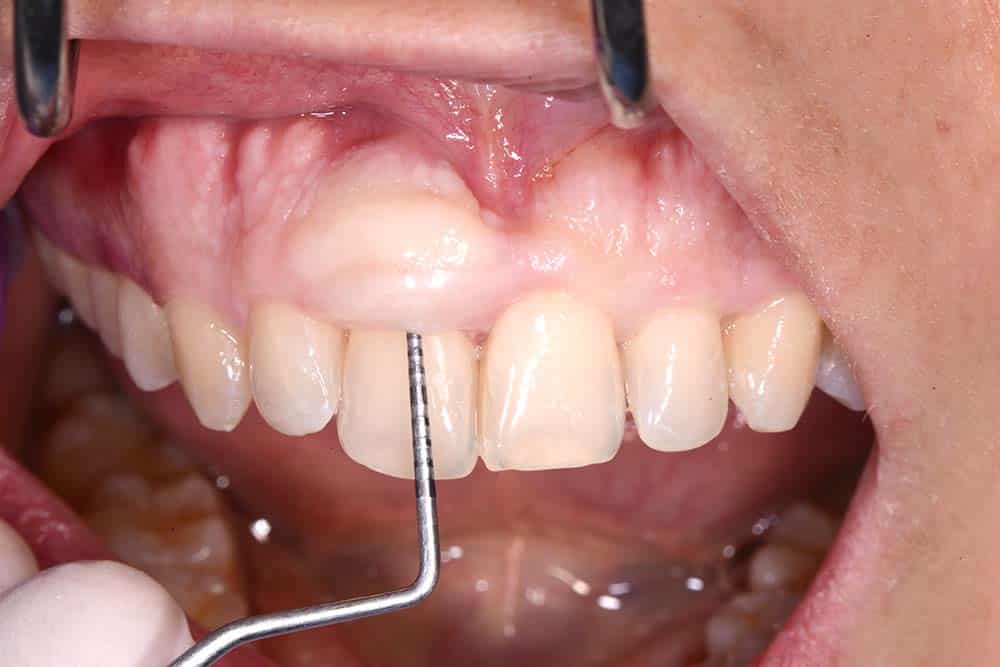
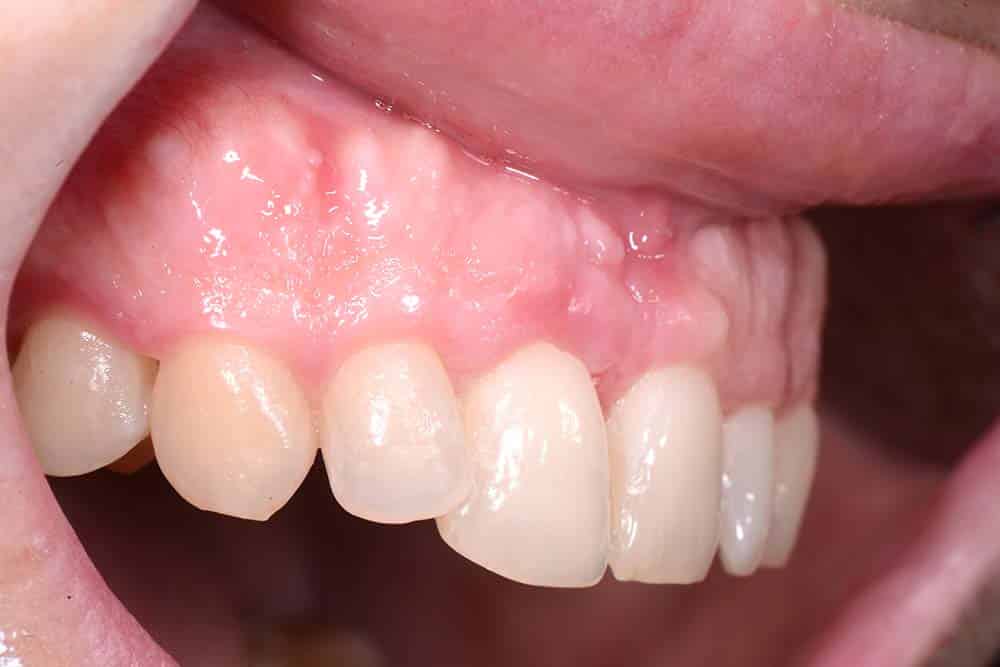
During the pandemic, the patient discontinued these hygiene sessions and returned for a check-up in 2023 with severe gingival inflammation, bone loss in the posterior regions, and an increase in gingival volume in the region of tooth 11.
Ultrasonic and sonic scalers were used in cleaning sessions to restore periodontal health.
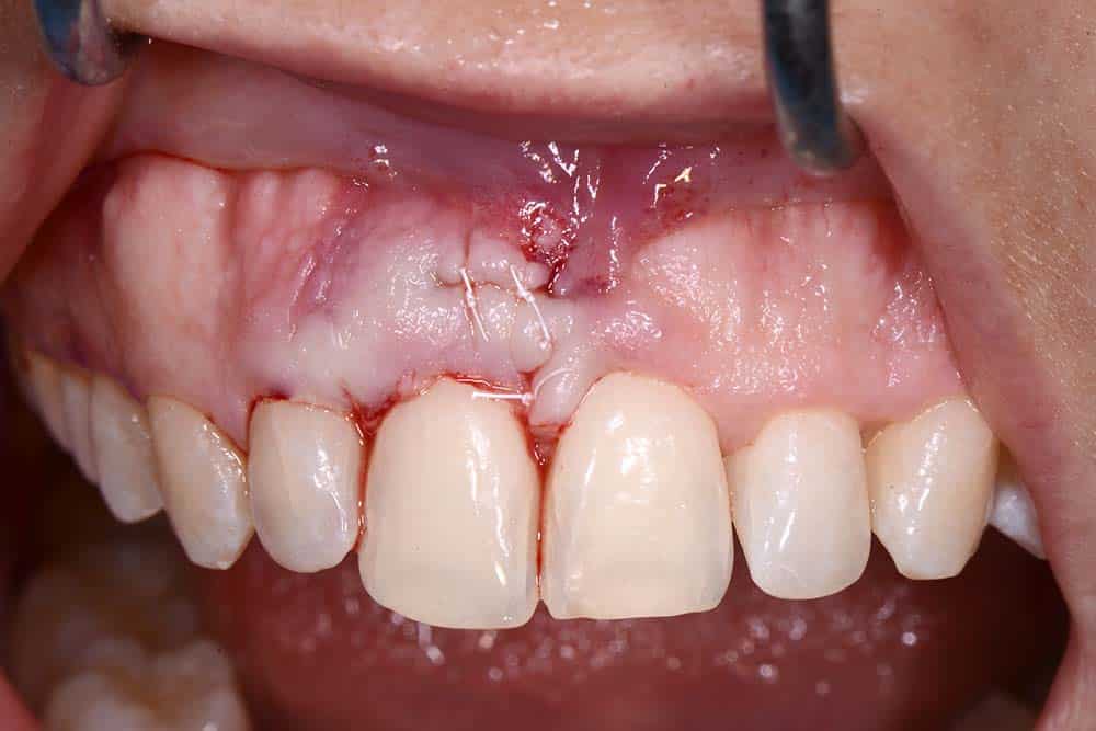
In 2024, a second surgical intervention has been performed to remove the excess soft tissue. A ball-shaped bur mounted on a sonic handpiece was used to eliminate the exostosis present in the regions of teeth 11 and 21.
The final follow-up is scheduled for 30 days post-surgery.

Gingival hypertrophy

Gingival hypertrophy – Occlusal view

Removal of Gingival hypertrophy with istological pickup
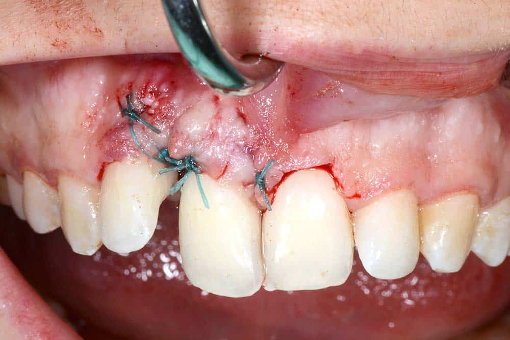
Suture
Healing
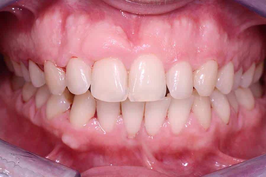
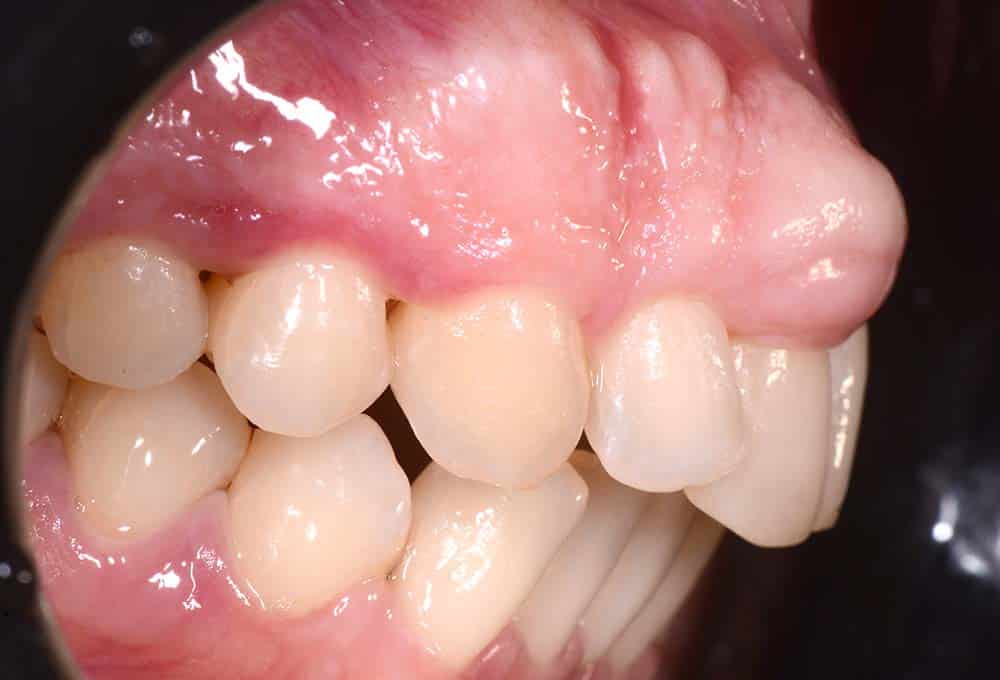
Bracketless fixed orthodontics treatment – Initial situation – Lateral view
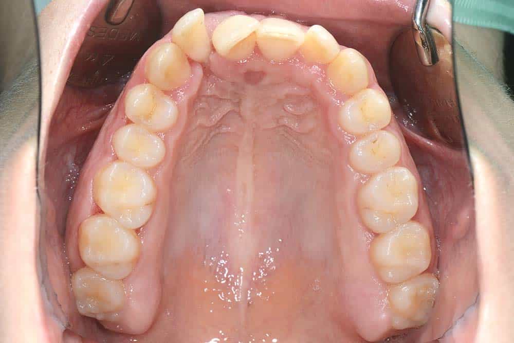
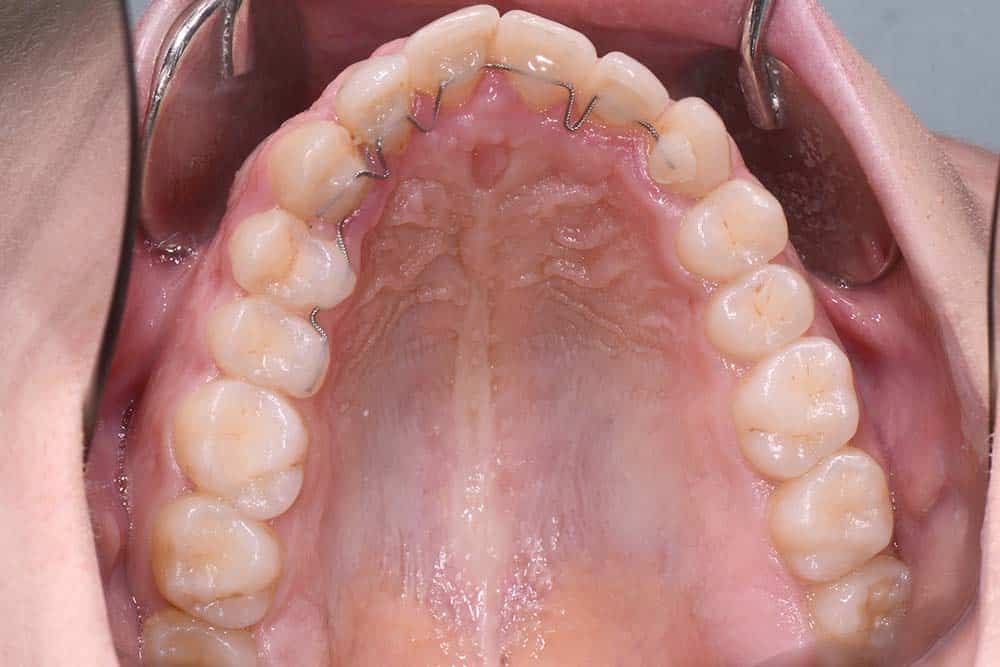
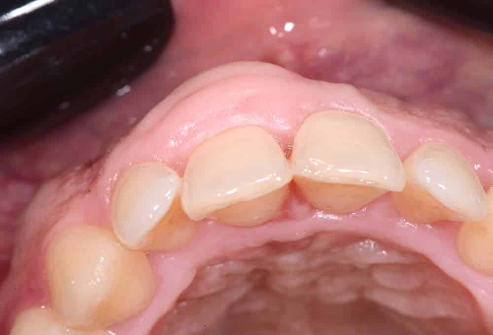
Bracketless fixed orthodontics treatment – Initial situation – Occlusal view upper arch
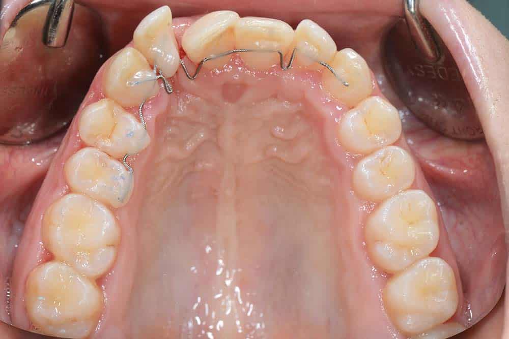
Bracketless fixed orthodontics treatment – First appliance – Occlusal view upper arch
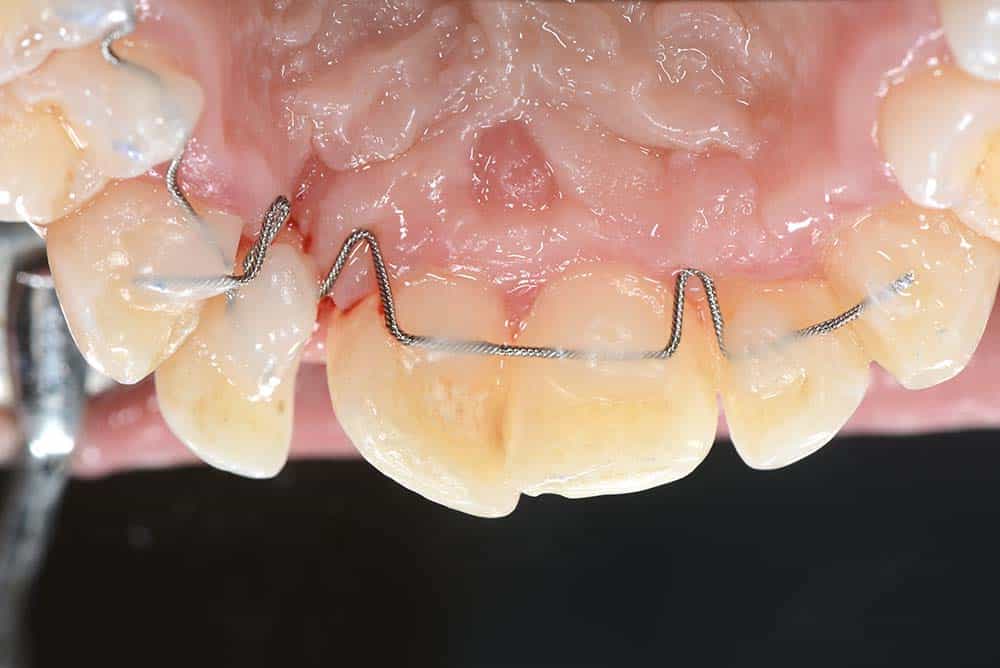
Bracketless fixed orthodontics treatment – First appliance – Occlusal view close up
Bracketless fixed orthodontics treatment – First appliance – Occlusal view close up
Bracketless fixed orthodontics treatment – Active retainer during the treatment
Bracketless fixed orthodontics treatment – Active retainer during the treatment
Bracketless fixed orthodontics treatment – Final result
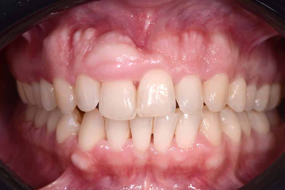
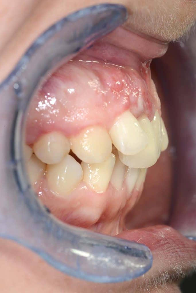
Gingival hypertrophy and Exostosis after 10 years
Gingival hypertrophy and Exostosis after 10 years – Frontal view
Gingival hypertrophy and Exostosis after 10 years – Occlusal view
Gingival hypertrophy and Exostosis after 10 years – Lateral view
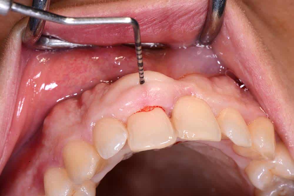
Probing length for the incision
Suture
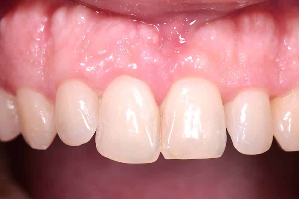
30 days Follow up
Follow up 30 days – Lateral view
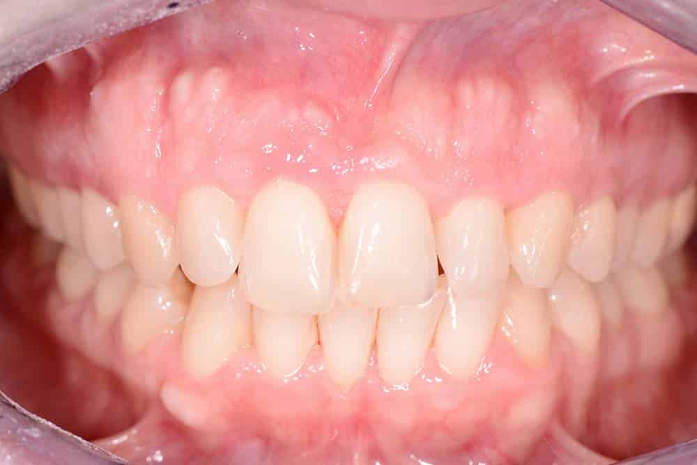
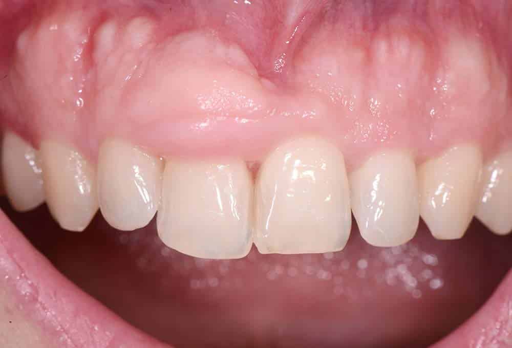
Final result
Video of “Removal of a gingival hypertrophy and reduction of an exostosis”
Share on: