Branch: Implantology, Oral Surgery.
Brands: Implanova Implant; ETK Implant, OneXeno graft.
Materials Used: Simple Lift Kit, osteotomes, dilators, sinus membrane separators, and xenograft.
Technique: Crestal sinus lift performed simultaneously with posterior implant placement; delayed implant placement; bite elevation; final prosthetic rehabilitation with a zirconia implant‑supported bridge.
A patient sought functional rehabilitation of the posterior maxilla. Clinical and CBCT examination revealed missing teeth 14, 15, and 16 with maxillary sinus pneumatization, leaving a residual bone height of 6.4 mm and adequate ridge width. A crestal sinus lift was performed using a non‑drilling Simple Lift kit, followed by implant placement.
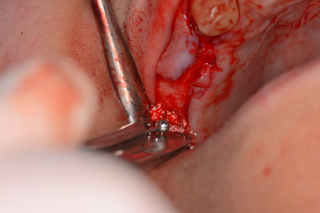
A trapezoidal flap was reflected followed by implant osteotomy for the anterior delayed implant using the ETK system, which is fully compatible with the Implanova implant connection. ETK was selected due to its aggressive thread design, providing enhanced primary stability.
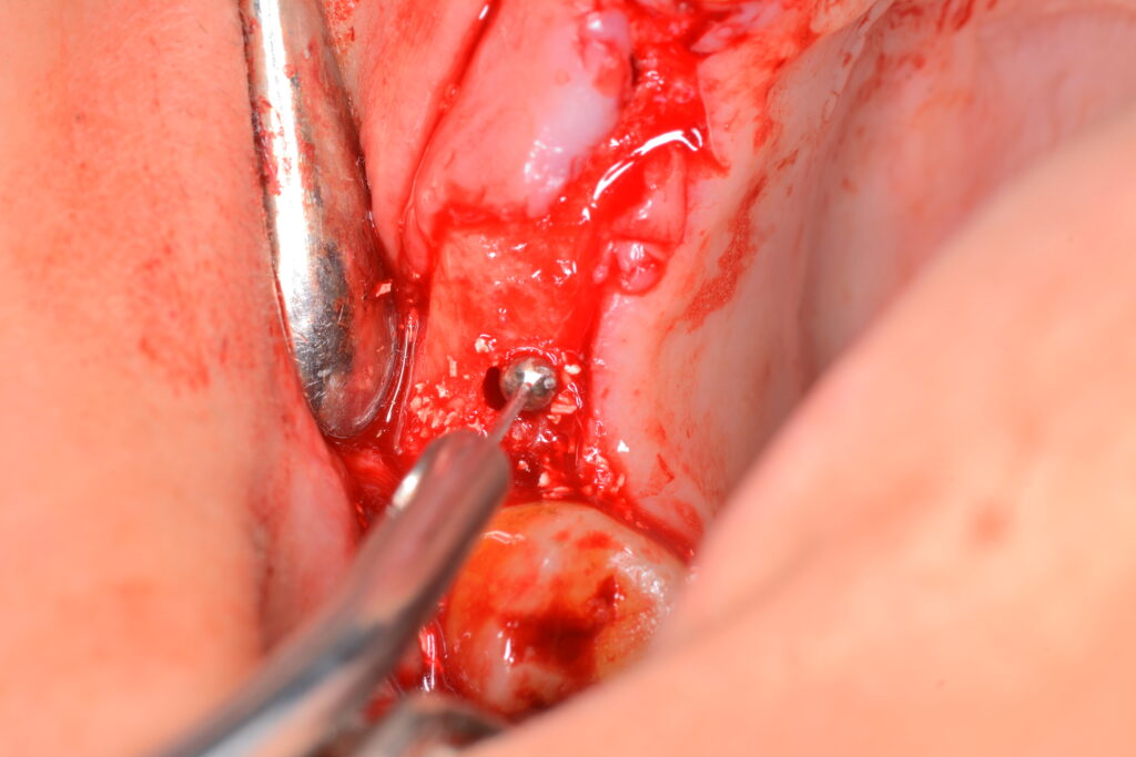
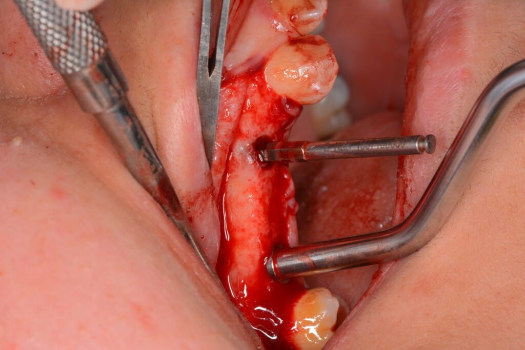
Posterior osteotomy was performed to 1 mm below the Schneiderian membrane. A modified osteotome (punch) was used to score the cortical bone beneath the membrane with gentle malleting. Circumferential color change from red to bluish hue indicated visualization of the Schneiderian membrane shadow. A separator was carefully used to elevate the membrane to the planned height. Sequential dilators with a gentle wiggling motion prepared the site to match the Implanova implant diameter; the blunt implant apex was advantageous in minimizing membrane perforation risk. Xenograft was placed, followed by implant placement.
During the three‑month healing period, the patient wore an RPD with a 2 mm bite elevation to facilitate neuromuscular adaptation. After three months, successful osseointegration and soft tissue healing were confirmed. A closed indirect impression was taken using coping and an analogue. Final rehabilitation was completed with a zirconia implant‑supported prosthesis, restoring the vertical dimension and achieving high patient satisfaction.
bibliography:
Effect of Different Crestal Sinus Lift Techniques for Implant Placement in the Posterior Maxilla of Deficient Height: A Randomized Clinical Trial
https://www.mdpi.com/2076-3417/13/11/6668
Evaluation of implant stability and increase in bone height in indirect sinus lift done with the osseodensification and osteotome technique: A systematic review and meta-analysis
https://www.thejpd.org/article/S0022-3913(23)00278-0/abstract
Comparative Evaluation of Survival Rate of Implant Placement Using Three Different Indirect Sinus Floor Elevation Techniques: A Systematic Review and Meta –Analysis
https://jchr.org/index.php/JCHR/article/view/8765
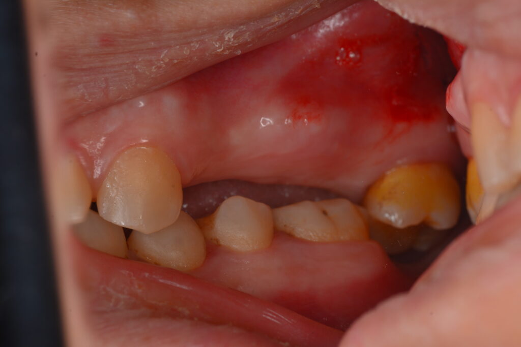
Preoperative lateral view demonstrating absence of teeth 14, 15, and 16 and inadequate interarch prosthetic space.
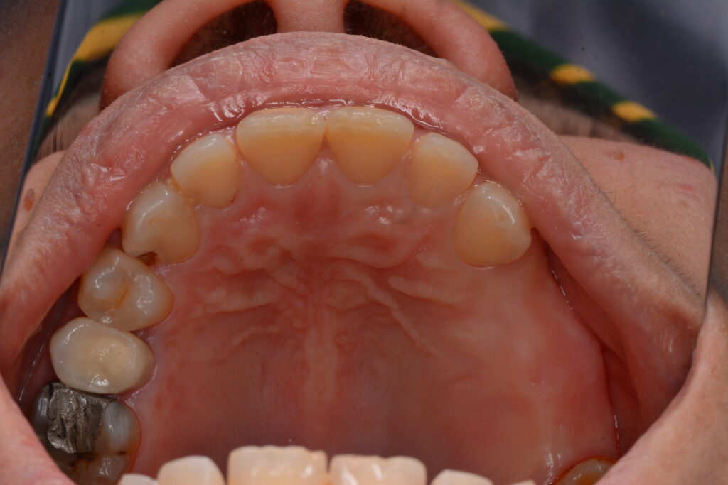
Preoperative occlusal view demonstrating the absence of teeth 14, 15, and 16.
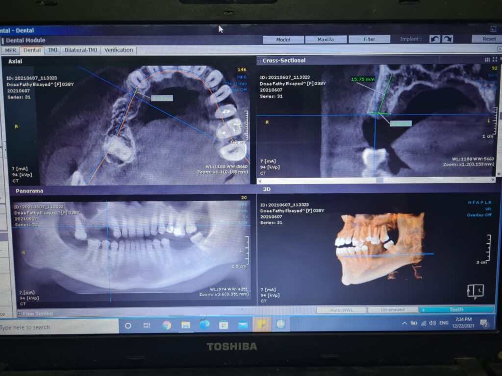
Preoperative CBCT analysis demonstrating adequate bone dimensions around the anterior planned implant, with a height of 15.7 mm and a width of 9.5 mm.
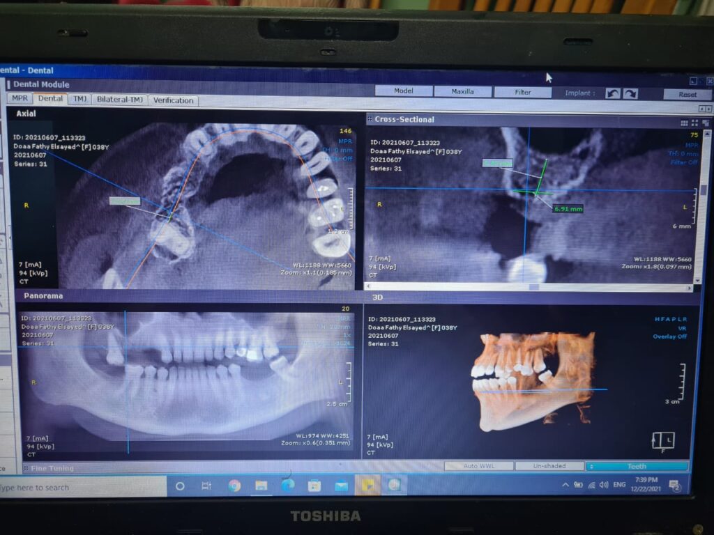
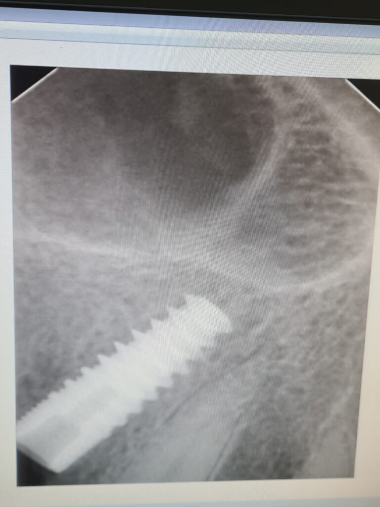
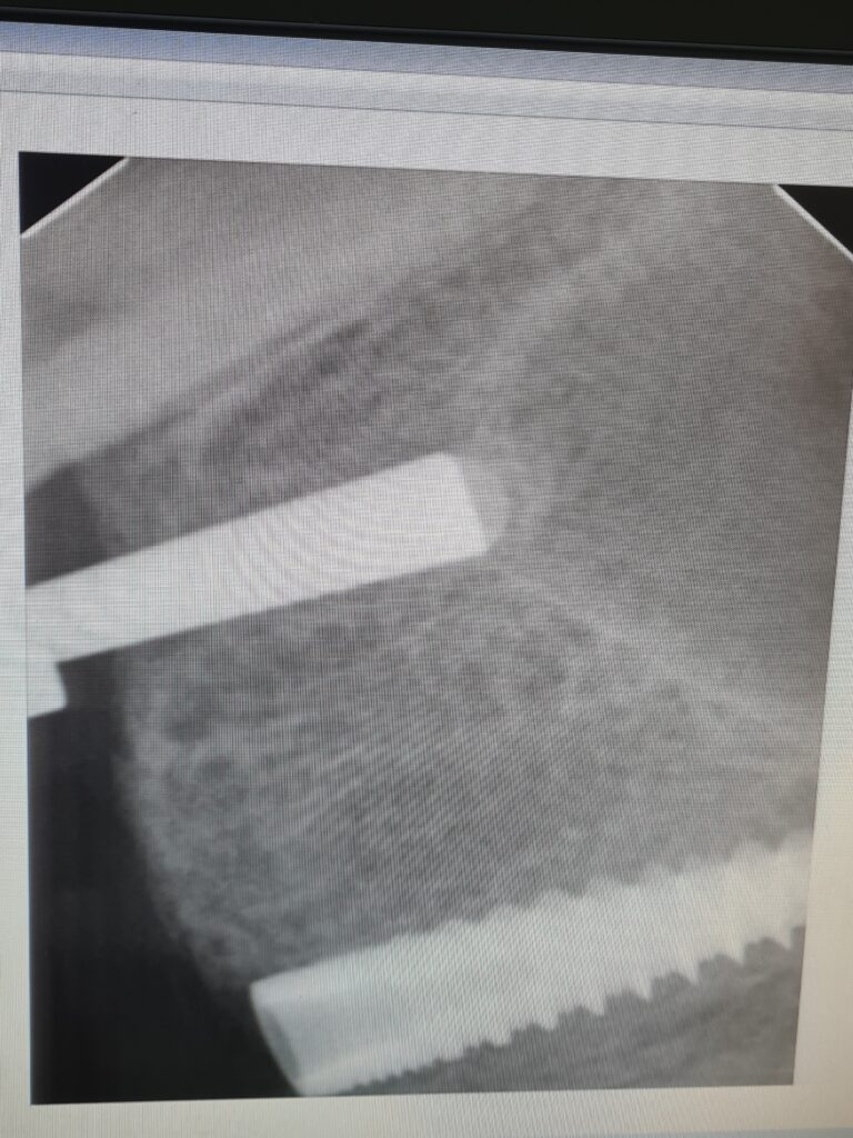
Preoperative CBCT analysis demonstrating insufficient bone height at the posterior planned implant site (6.4 mm) with adequate ridge width (7 mm).
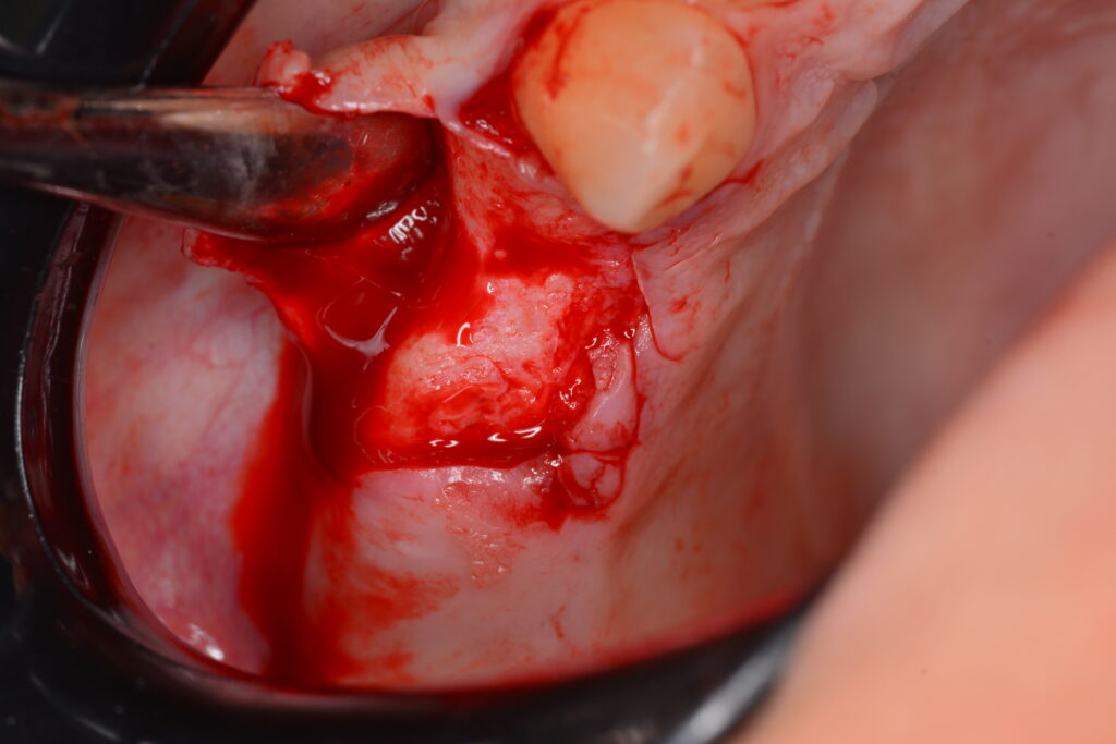
Trapezoidal flap design
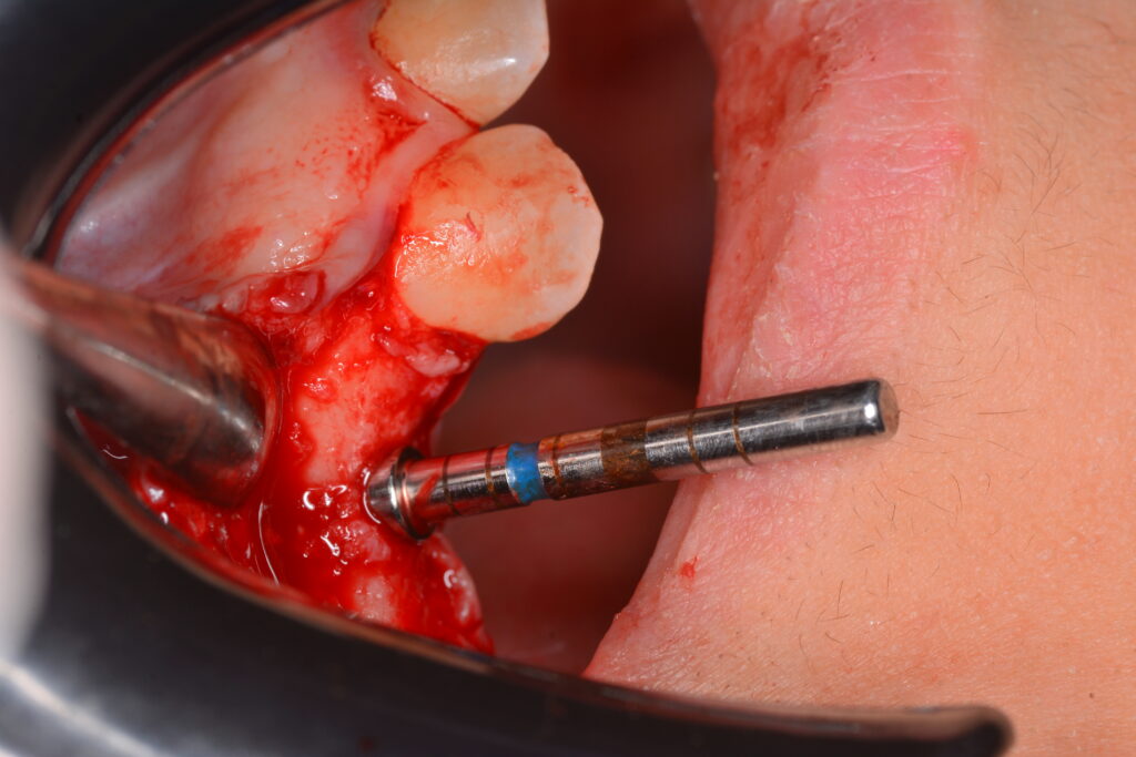
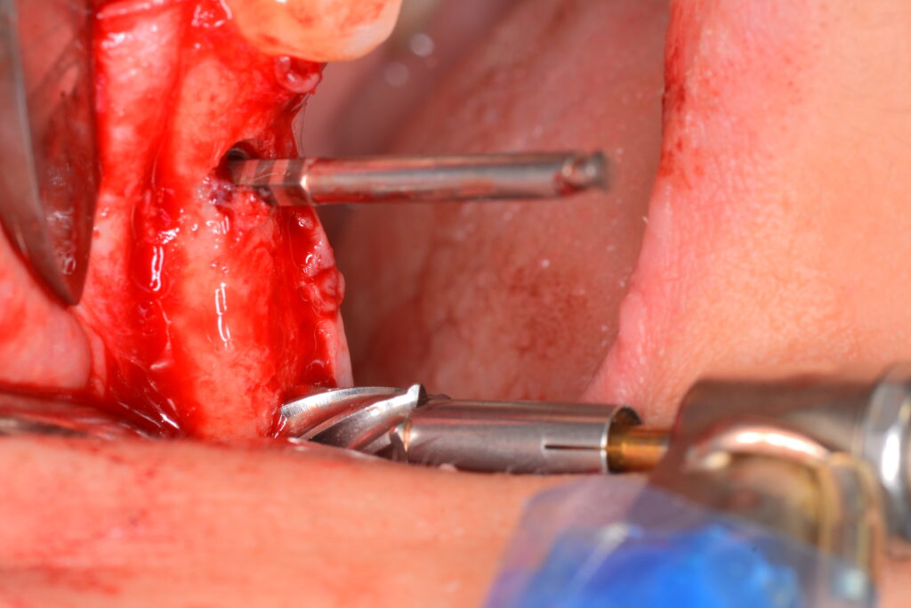
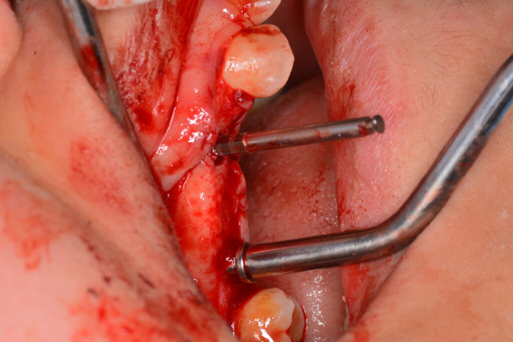
Paralleling pin confirming proper osteotomy angulation.
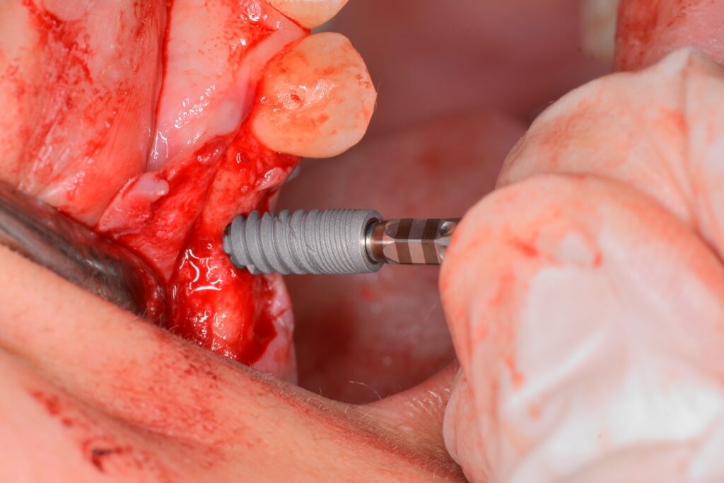
ETK implant placement (Side view)
ETK implant placement (occlusal view)
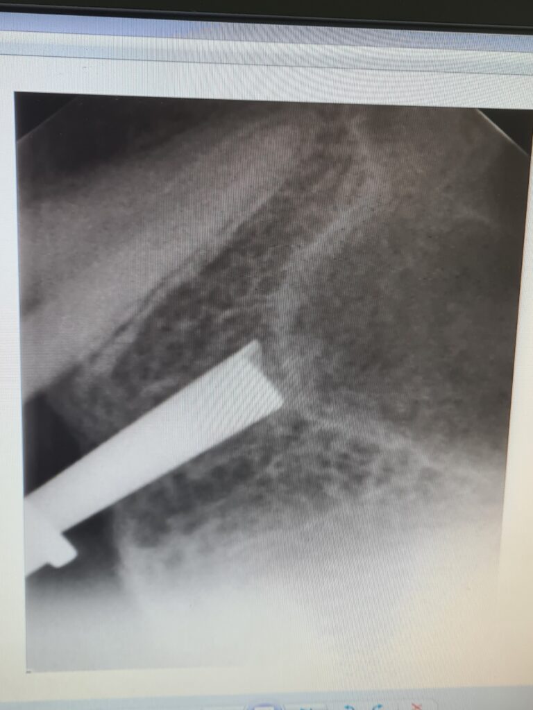
Immediate post-implant X-ray.
Posterior drilling performed to a depth of 1 mm below the sinus floor.
demarking of the sinus floor using a punch and mallet from the SimpleLift kit.
The punch before the tapping.
After sinus floor decortication.
Xenograft in place.
Xenograft in place.
Sequential dilation of the osteotomy to match the corresponding Implanova implant diameter, with simultaneous apical condensation of the xenograft.
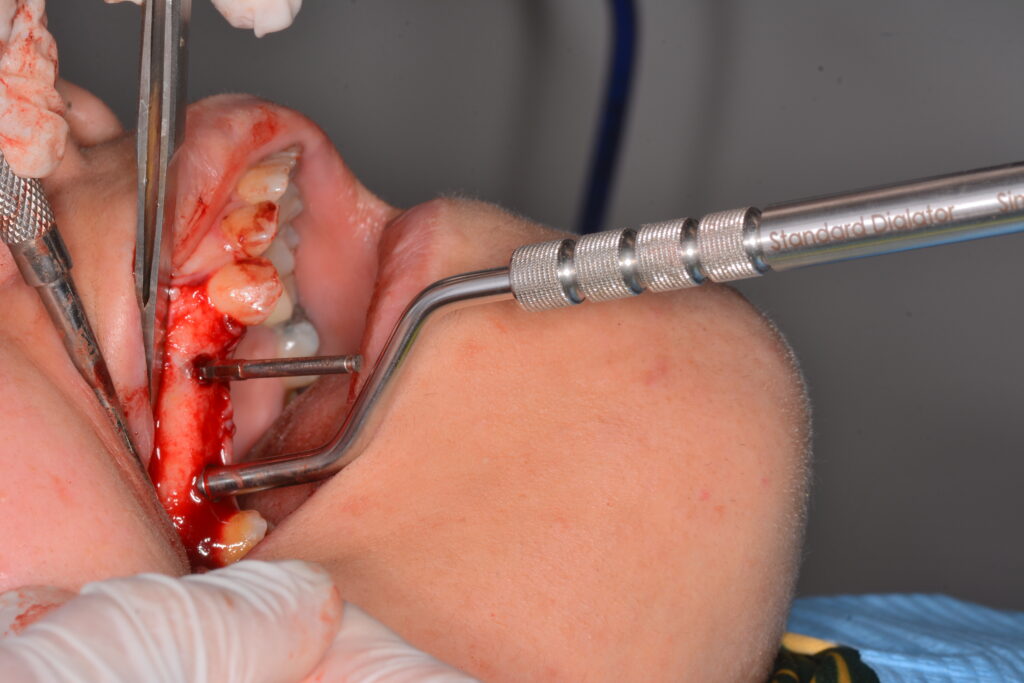
Sequential dilation of the osteotomy to match the corresponding Implanova implant diameter, with simultaneous apical condensation of the xenograft.
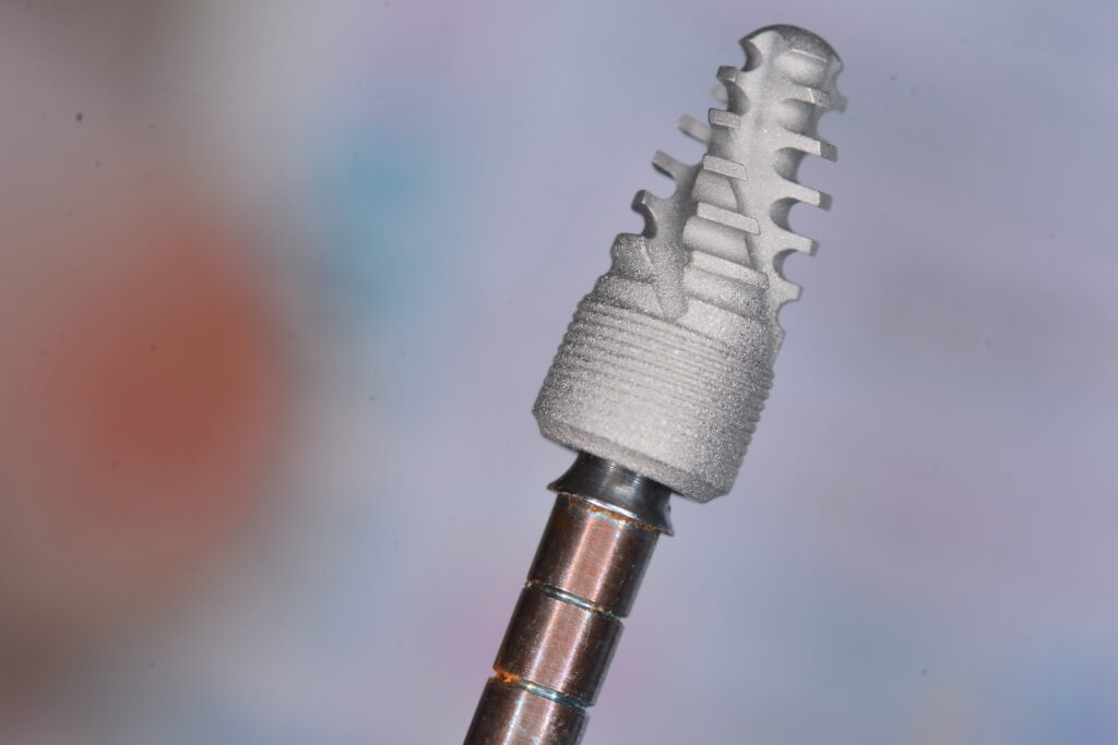
Implanova implant fixture showing grafting channels and a blunt apex to reduce the risk of injury to adjacent anatomical structures.
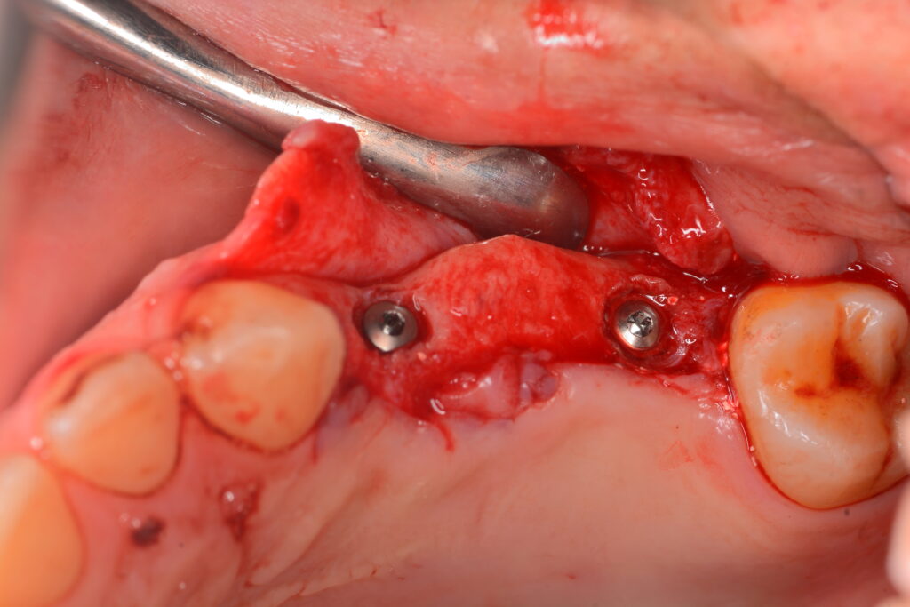
Implant placing (lateral view)
Implant placing (occlusal view)
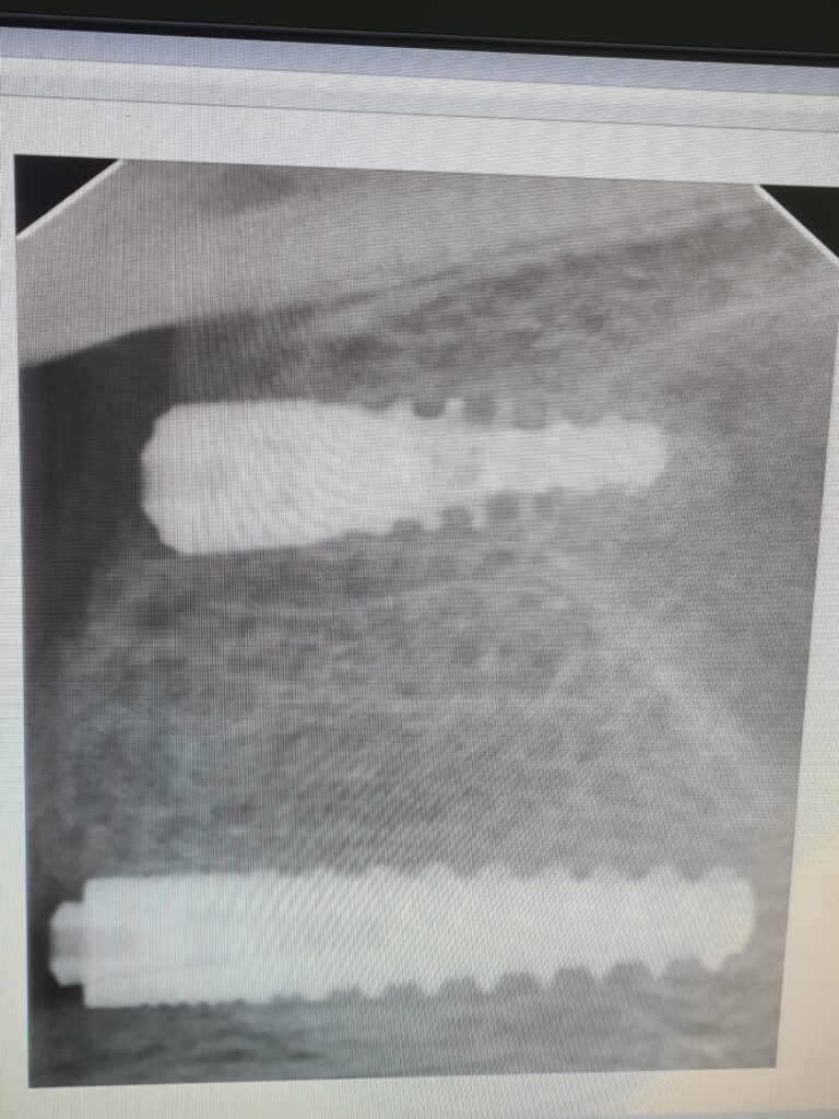
Immediate postoperative X‑ray showing a dome‑shaped sinus lift and favorable parallel alignment of the two implants
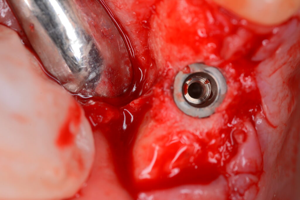
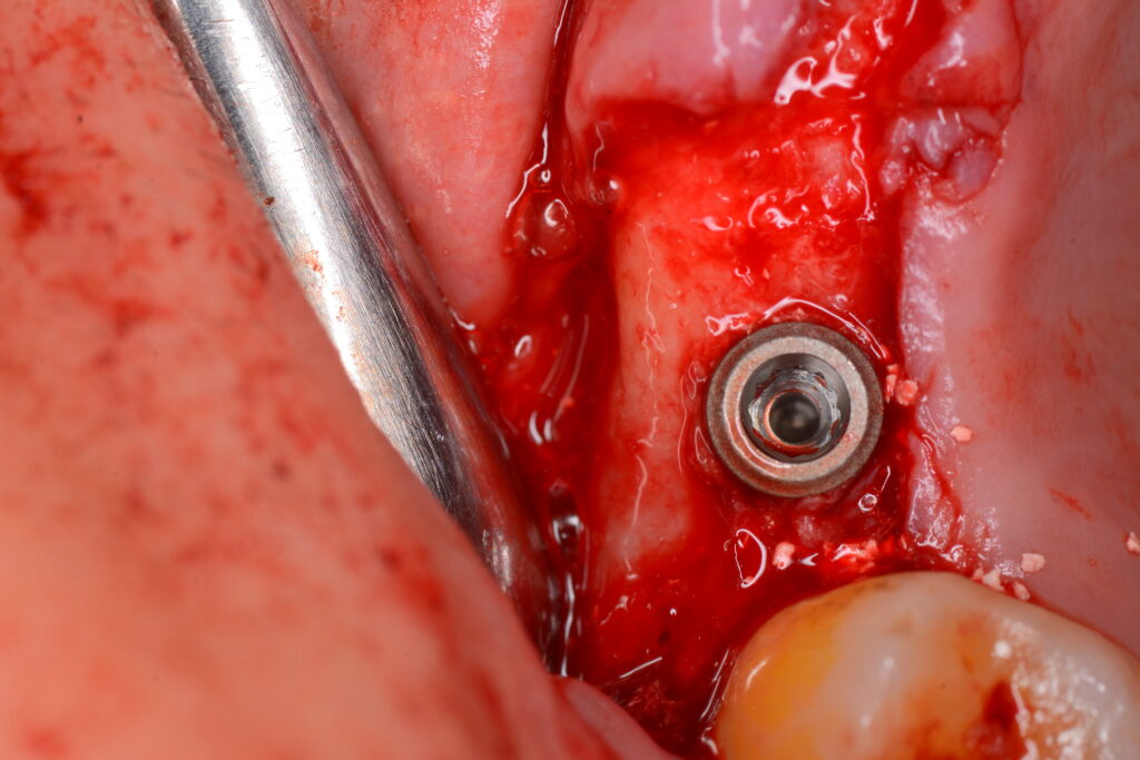
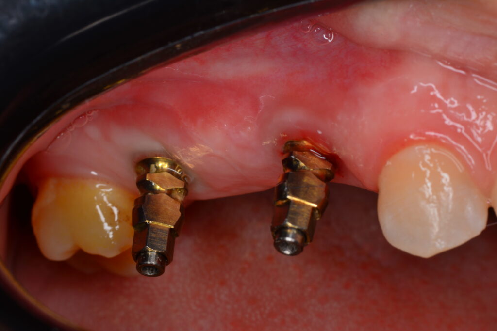
Postoperative implant placement (occlusal view)..
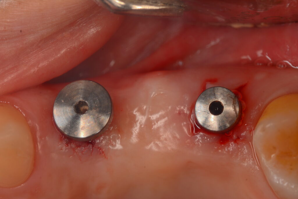
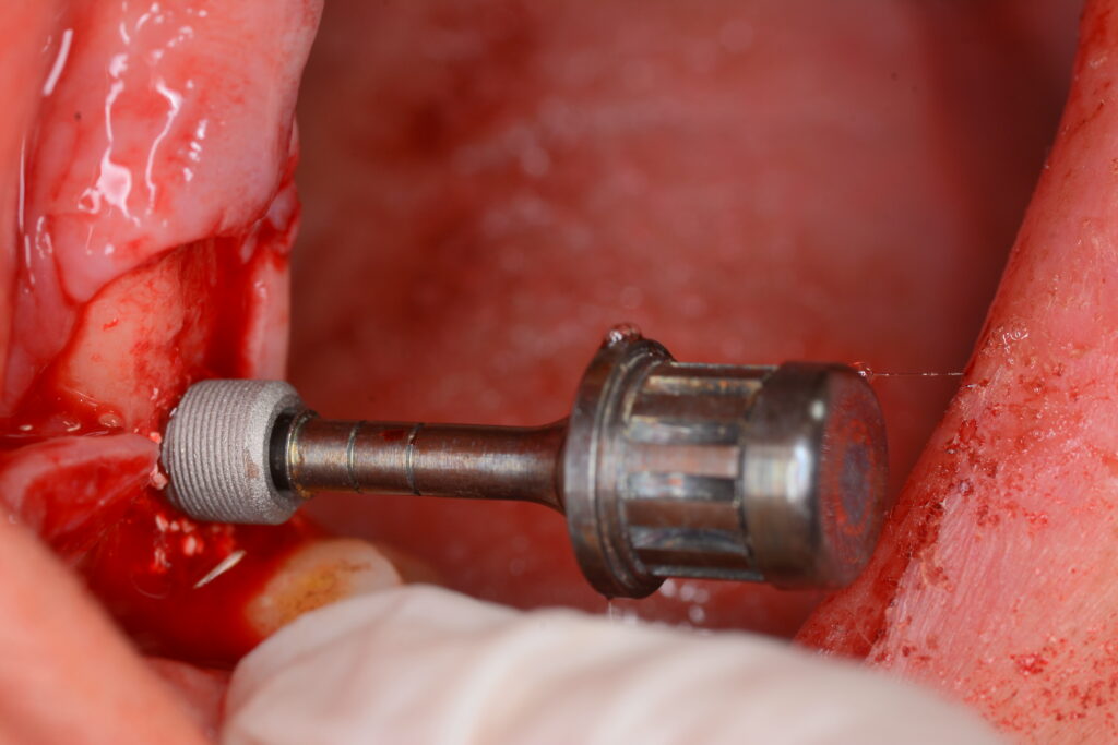
Healing abutments in place.
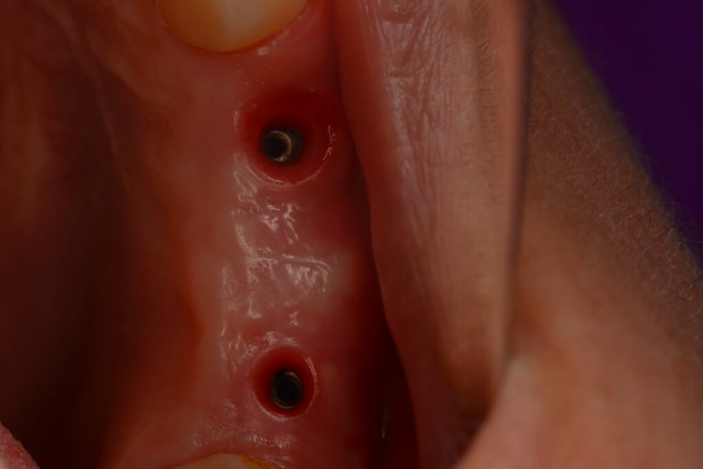
Soft tissue healing after 3 months.
Indirect impression technique (closed tray)
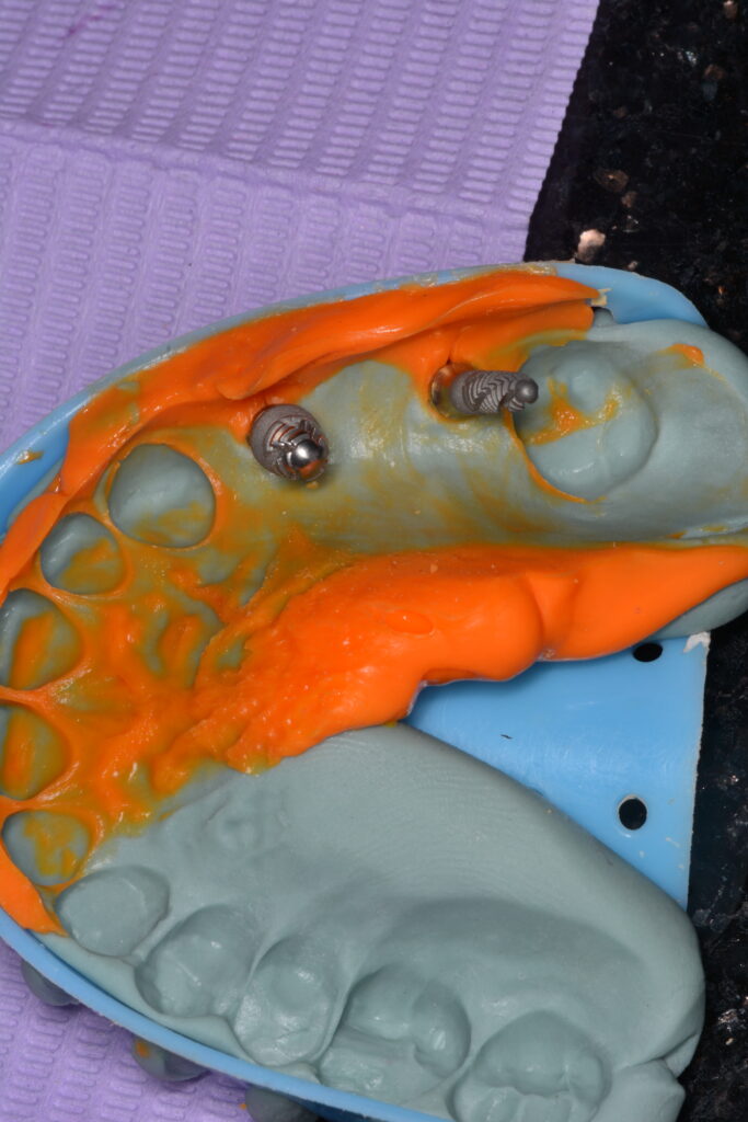
Indirect impression technique (closed tray)
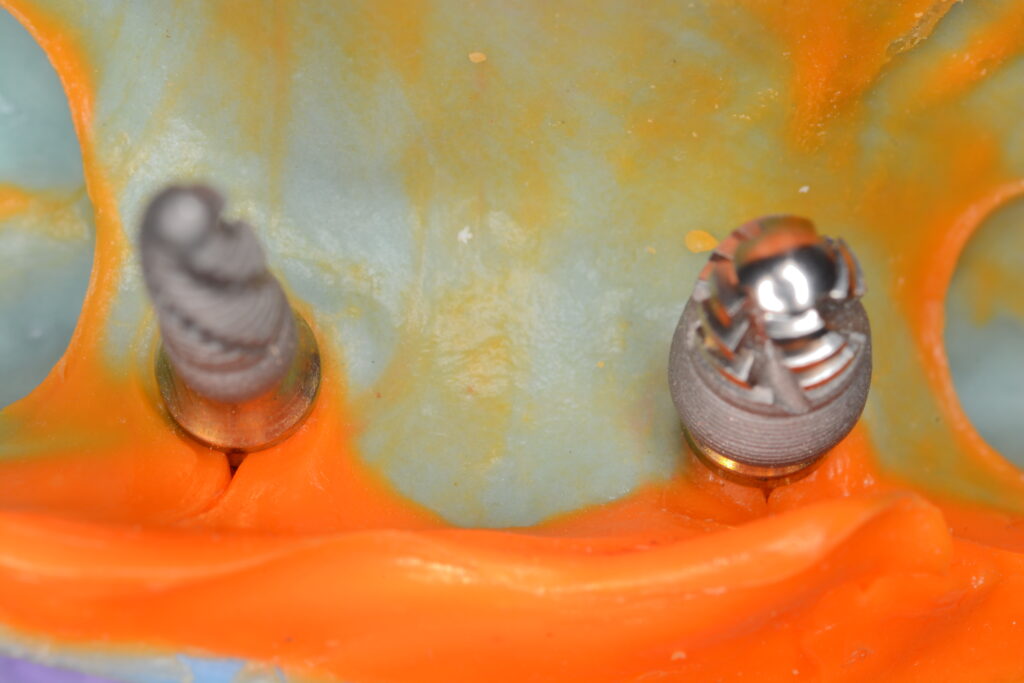
Indirect impression technique (closed tray)
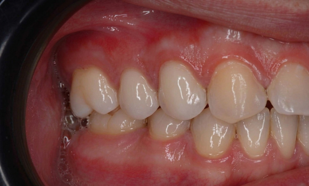
Final zirconia implant‑supported prosthesis showing proper occlusion and satisfactory functional outcome.
Share on: