Microscope-Assisted Root Canal Treatment of Maxillary Second Molar (#7) with Bioceramic Sealer
Dr Hamza Zahid
Biomimetic & Adhesive Dentistry
Dr Hamza Dental Center – Lahore, Pakistan
ABSTRACT
This clinical report demonstrates the management of a maxillary second molar (#7) with intricate canal anatomy using a dental operating microscope and rubber dam isolation. Enhanced magnification permitted precise canal negotiation, efficient irrigation activation and controlled obturation. A bioceramic sealer was selected for its exceptional sealing ability, dimensional stability and bioactive stimulation of periapical healing. The case highlights how advanced visual control and biomimetic endodontic materials optimise biological and restorative outcomes.
CLINICAL REPORT
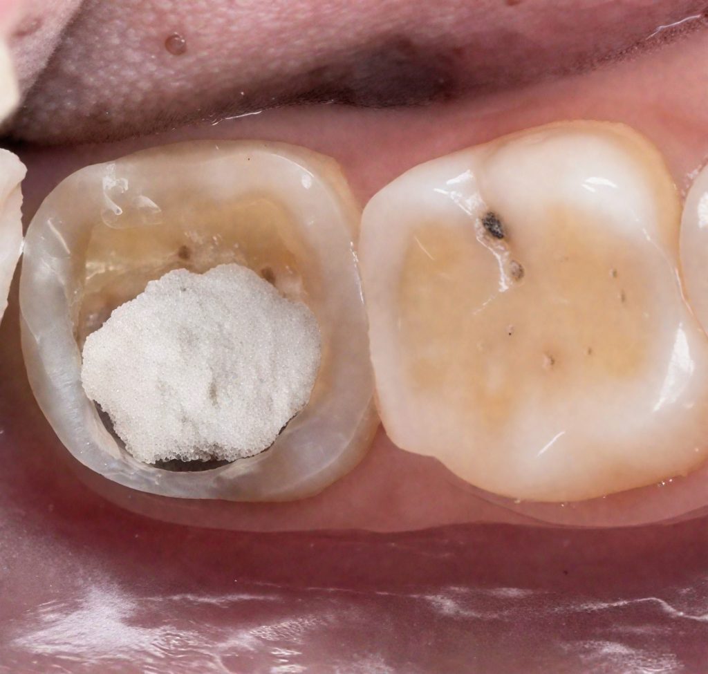
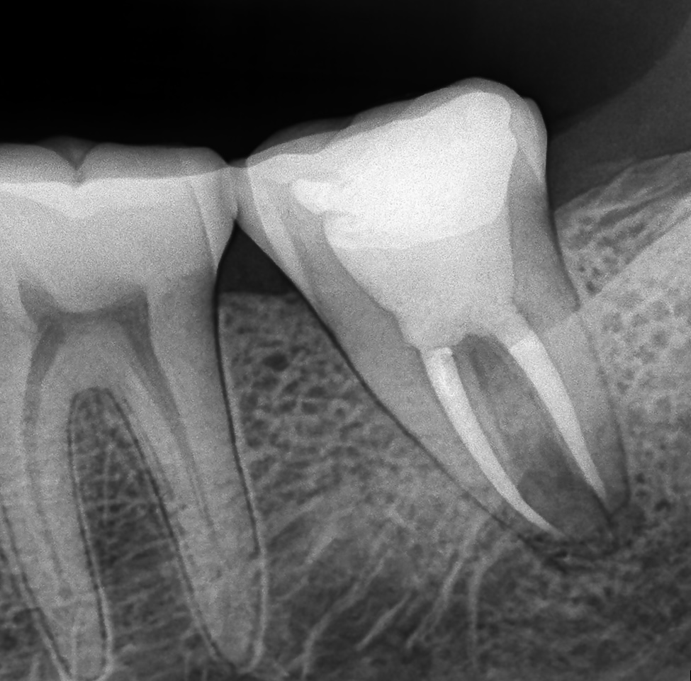
The patient presented with discomfort on biting and a history of intermittent dull pain on the upper left posterior side. Radiographic examination revealed previously untreated carious involvement extending into the pulp chamber of tooth #7.
Isolation and Access
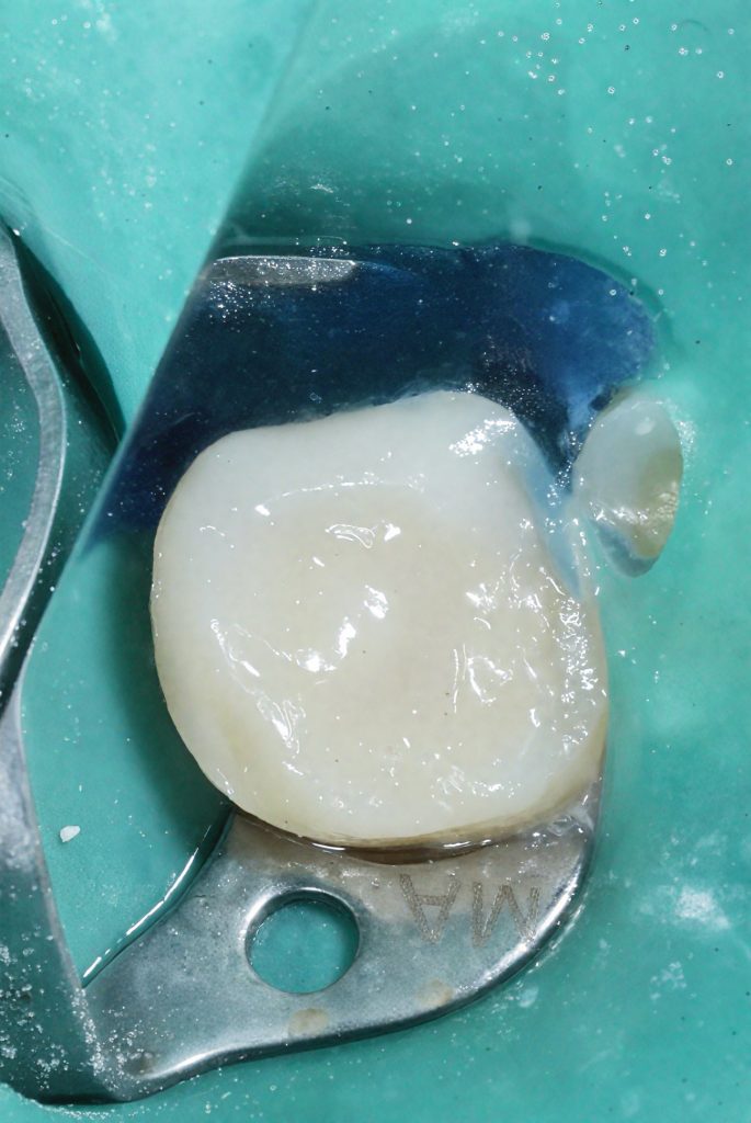
After administration of local anaesthesia, rubber dam isolation was achieved to ensure aseptic conditions. Under microscope magnification, the pulp chamber was accessed conservatively, maintaining maximum structural integrity.
Canal Negotiation and Instrumentation
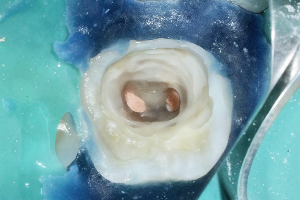
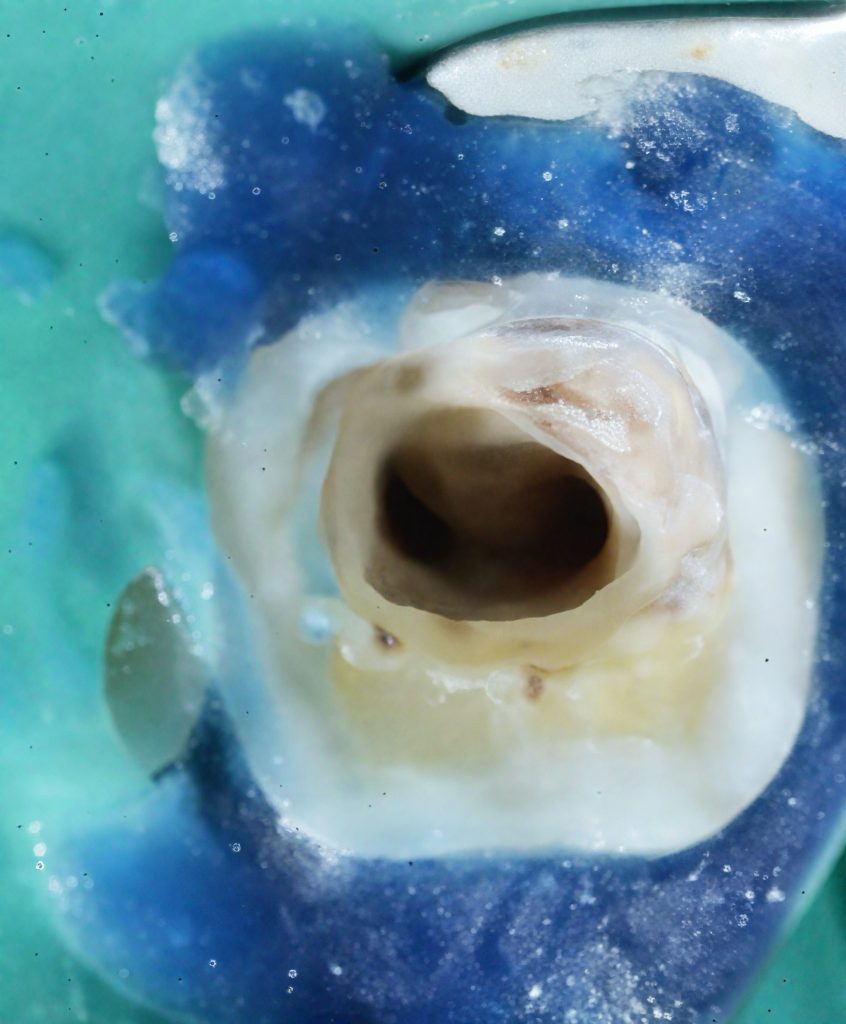
All canals were located and verified under high magnification. Glide-path creation was performed using stainless-steel hand files, followed by rotary NiTi shaping. The microscope facilitated clear identification of the canal orifices, prevention of ledging and maintenance of original curvature.
Irrigation Protocol
The canals were irrigated with 5.25 % sodium hypochlorite, followed by 17 % EDTA, both ultrasonically activated for improved penetration and debris removal. Final rinse with distilled water ensured neutralisation before obturation.
Obturation
Obturation was achieved with a single-cone technique using bioceramic sealer to obtain a three-dimensional fill. The material’s bioactivity encouraged the formation of hydroxyapatite at the sealer–dentine interface, enhancing long-term sealing and periapical repair.
Coronal Seal
A composite coronal build-up was completed immediately after obturation to prevent reinfection and reinforce the cuspal structure.
Outcome
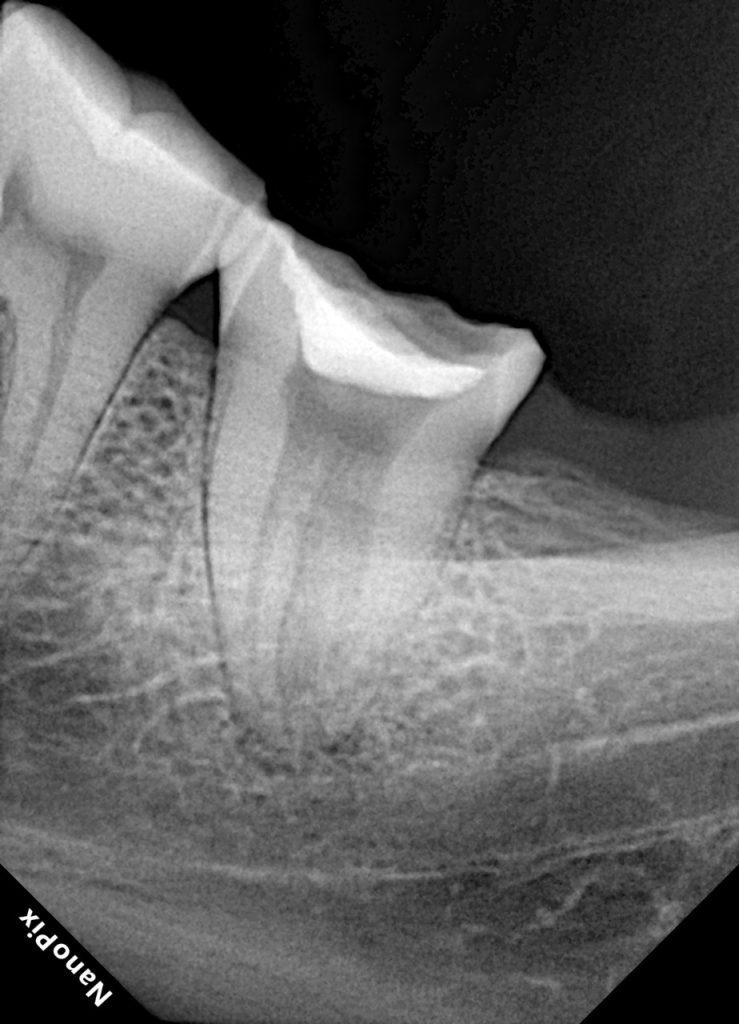
Post-operative radiographs demonstrated excellent obturation density and apical seal. The patient remained asymptomatic at review, with periapical healing evident radiographically after follow-up.
DISCUSSION
Microscope-assisted endodontics represents the modern standard of precision. Magnification not only enhances identification of canal morphology but also allows clinicians to perform minimally invasive shaping and thorough debridement.
The integration of bioceramic sealers—notably their bioactive potential and chemical adhesion to dentine—further supports periapical regeneration and reduces microleakage compared with traditional resin or zinc-oxide–eugenol sealers.
In this case, the synergy of magnification, isolation, and biomimetic obturation produced a predictable, biologically sound outcome.
CONCLUSION
The successful treatment of a complex posterior tooth under the microscope demonstrates how visual precision and bioactive materials elevate both the biological and mechanical reliability of root canal therapy. Proper isolation, controlled irrigation and the use of bioceramic sealers ensure long-term periapical health and coronal integrity.
FIGURE REFERENCES
- Fig 1. Pre-operative radiograph showing carious involvement.
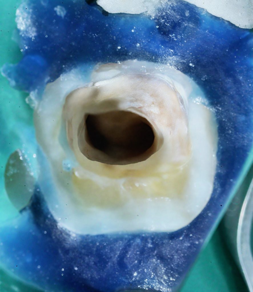
- Fig 2. Rubber dam isolation and access cavity under microscope.
- Fig 3. Canal negotiation and instrumentation.
- Fig 4. Obturation with bioceramic sealer.
- Fig 5. Post-operative radiograph demonstrating apical seal.
BIBLIOGRAPHY
- Torabinejad M, Parirokh M. Mineral trioxide aggregate: a comprehensive literature review—Part II: leakage and biocompatibility. J Endod. 2010;36(2):190-202.
- Zhang W, Li Z, Peng B. Assessment of a new root canal sealer’s apical sealing ability and cytotoxicity. Dent Mater J. 2009;28(5):524-528.
- de Deus G et al. Sealing ability of three root canal sealers using a fluid filtration model. Int Endod J. 2007;40(10):844-852.
- Kim S, Krieger E. Microscope-enhanced endodontics: the state of the art. Dent Clin North Am. 2010;54(2):391-413.
{"source_type":"hypic","tiktok_developers_3p_anchor_params":"{"template_id":"","client_key":"awgvo7gzpeas2ho6","filter_id":[],"capability_extra_v2":{"edit":[{"panel":"hd_quality_picture"},{"panel":"auto_edit_pro"},{"panel":"smart_color_button"}]},"capability_key":["edit"]}","data":{"os":"ios","enter_from":"enter_launch","imageEffectId":"","stickerId":"","playId":"","filterId":"","activityName":"","infoStickerId":"","appversion":"7.4.0","product":"retouch","pictureId":"FEC36F85-E2C3-4B2B-90F4-F124D7916D2F"}}
{"tiktok_developers_3p_anchor_params":"{"template_id":"","client_key":"awgvo7gzpeas2ho6","filter_id":[],"capability_extra_v2":{"edit":[{"panel":"hd_quality_picture"},{"panel":"auto_edit_pro"}]},"capability_key":["edit"]}","data":{"activityName":"","os":"ios","stickerId":"","playId":"","imageEffectId":"","appversion":"7.4.0","pictureId":"80FBDF67-C028-47FC-8E3C-5321988C68DF","product":"retouch","enter_from":"enter_launch","filterId":"","infoStickerId":""},"source_type":"hypic"}
{"tiktok_developers_3p_anchor_params":"{"template_id":"","capability_extra_v2":{"edit":[{"panel":"hd_quality_picture"},{"panel":"auto_edit_pro"}]},"capability_key":["edit"],"client_key":"awgvo7gzpeas2ho6","filter_id":[]}","data":{"stickerId":"","pictureId":"C1121DCE-96F0-41CA-AC2D-DB197FB36451","os":"ios","appversion":"7.4.0","playId":"","product":"retouch","enter_from":"enter_launch","infoStickerId":"","filterId":"","activityName":"","imageEffectId":""},"source_type":"hypic"}
{"data":{"imageEffectId":"","stickerId":"","os":"ios","appversion":"7.4.0","filterId":"","infoStickerId":"","playId":"","enter_from":"enter_launch","activityName":"","pictureId":"3C3F2496-8990-435D-9B96-13A6A1739242","product":"retouch"},"source_type":"hypic","tiktok_developers_3p_anchor_params":"{"client_key":"awgvo7gzpeas2ho6","capability_key":["edit"],"template_id":"","capability_extra_v2":{"edit":[{"panel":"hd_quality_picture"},{"panel":"auto_edit_pro"}]},"filter_id":[]}"}
{"tiktok_developers_3p_anchor_params":"{"template_id":"","filter_id":[],"capability_key":["edit"],"capability_extra_v2":{"edit":[{"panel":"hd_quality_picture"},{"panel":"smart_color_button"}]},"client_key":"awgvo7gzpeas2ho6"}","data":{"enter_from":"enter_launch","product":"retouch","stickerId":"","infoStickerId":"","filterId":"","activityName":"","playId":"","appversion":"7.4.0","imageEffectId":"","os":"ios","pictureId":"BC1506AA-7DF9-4D7A-A4A3-2561C40BACDC"},"source_type":"hypic"}
Share on: