Gingival Recession treatment FGG
1- Type 2 gingival recession with interproximal CAL loss.
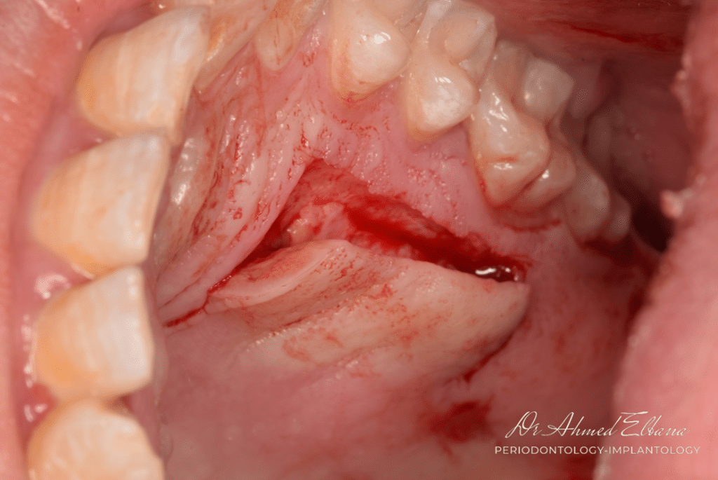
2- Recipient bed preparation.
3- Harvesting FGG.
4- Graft adaptation.
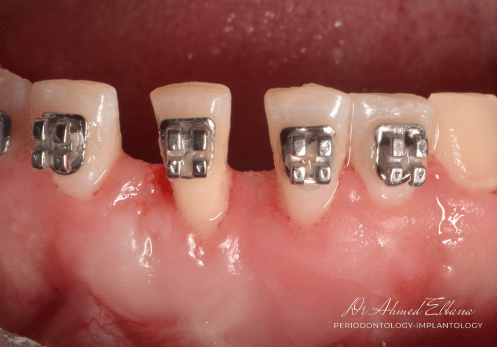
5- Orthodontic treatment
6- Follow up

this figure shows:
- gingival recession with root surface exposure.
-lack of attached keratinized gingiva.
-high frenulum pull.

using 15c scalpel blade horizontal incision at the level of MGJ extended to involve the defect. partial thickness flap preparation is performed to displace the muscle fibers in apical direction away from the denuded root surface.
FGG was harvested from the palate at the premolar region. partial thickness palatal graft was adequate for the treatment purpose.
the palate was sutured using collagen sponge and cross mattress sutures.
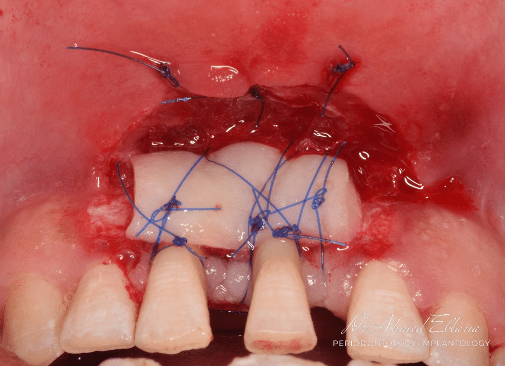
perfect adaptation is essential for plasmic diffusion which is mandatory for graft integration.
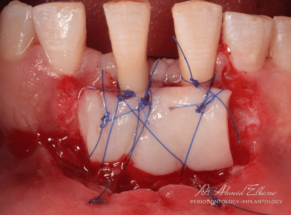
the size of the graft is preferred to be smaller than the recipient bed to ensure vascularization into the graft from all directions
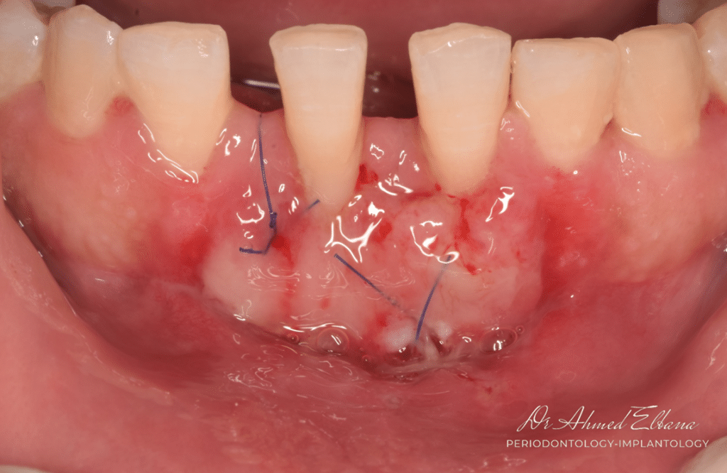
two weeks post operative. the figure show new capillaries and revascularization of the graft.

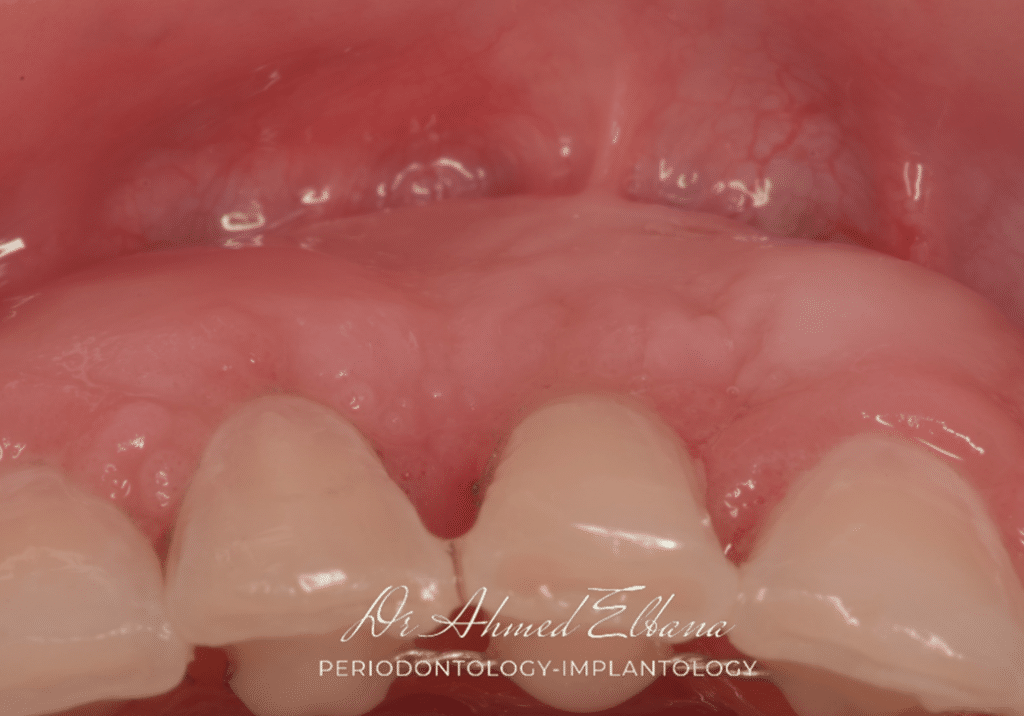
the figure shows perfect tissue integration and color matching.
note tissue thickness after healing and maturation.

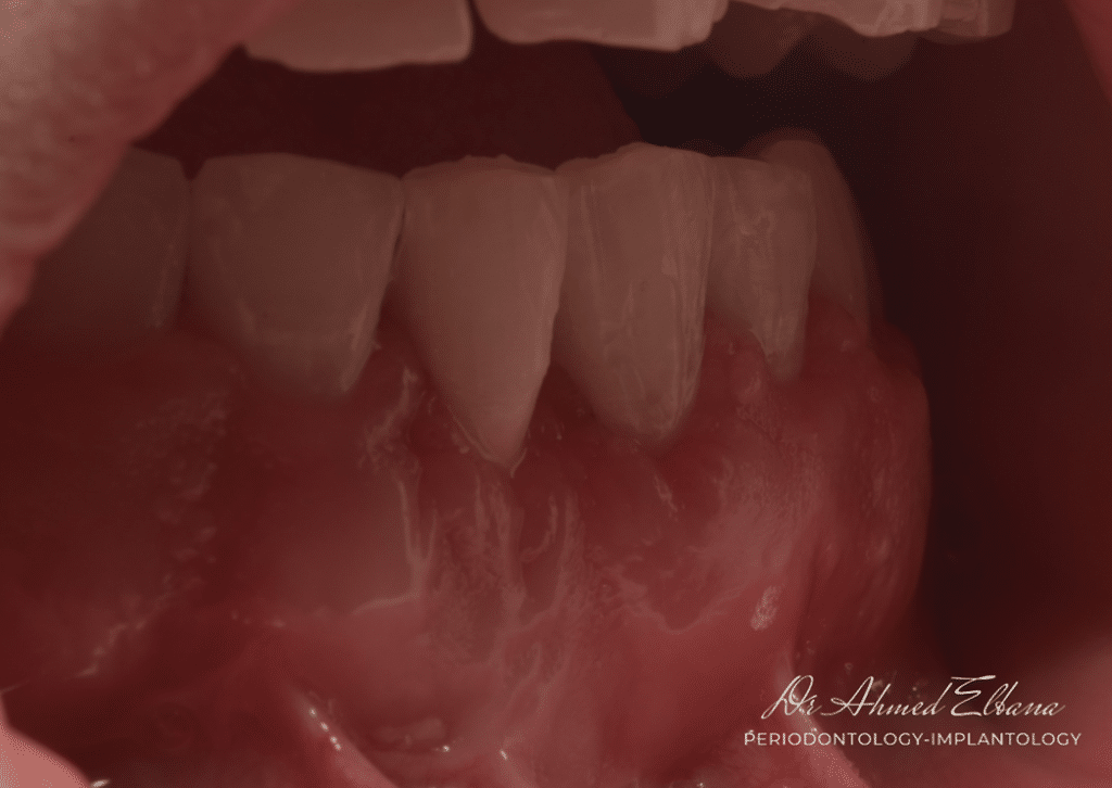
occlusal view to show tissue thickness formed after graft maturation.
two years follow up period
Share on: