Microscope-Assisted Re-Endodontic Management of a Molar with Broken Instrument, Ledge Bypass, and MTA Repair Followed by Biomimetic Cuspal Coverage Restoration
Abstract (≈480 characters)
This case presents the retreatment of a molar with a fractured instrument and canal ledge. Under microscope guidance, the broken orifice opener was ultrasonically removed, the ledge bypassed, and the perforation sealed with MTA. The canal system was then obturated using a bioceramic sealer. A definitive biomimetic restoration was performed using Deep Marginal Elevation (DME), GC EverX Flow, and Tokuyama Estelite Sigma Quick for optimal reinforcement and esthetics.
Author CV
Dr Hamza Zahid, BDS
Microscopic Restorative & Cosmetic Dentist
CEO – Dr Hamza Dental Center, Lahore (Pakistan)
Focus Areas – Micro-Endodontics | Instrument Retrieval | MTA Repair | Biomimetic Posterior Restorations
Clinical Workflow
1️⃣ Pre-operative Assessment
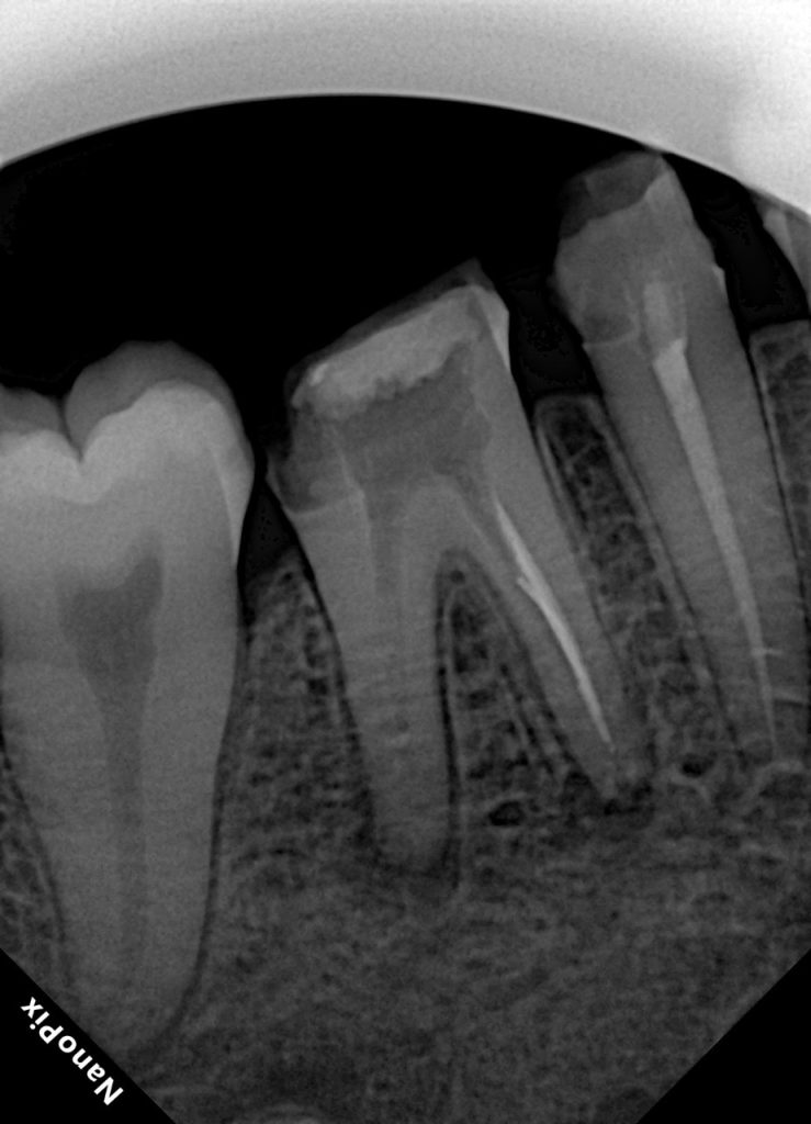
Patient presented with persistent dull pain in a previously treated mandibular molar (Fig 1). Radiograph revealed under-filled canals, apical radiolucency, and a fractured orifice opener lodged in the distal canal. Diagnosis: failed RCT with symptomatic periapical periodontitis.
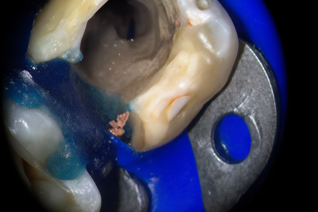
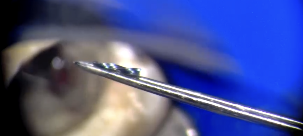
2️⃣ Access and Microscope Exploration
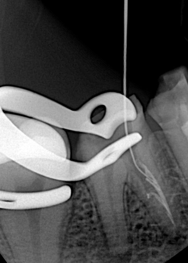
Rubber-dam isolation achieved. Conservative access refined under a dental microscope (Fig 2). The broken instrument was visualized in the coronal third of the distal canal. Ultrasonic tips (ET18D and ProUltra Endo) were used to trephine dentin around the fragment and safely dislodge it.
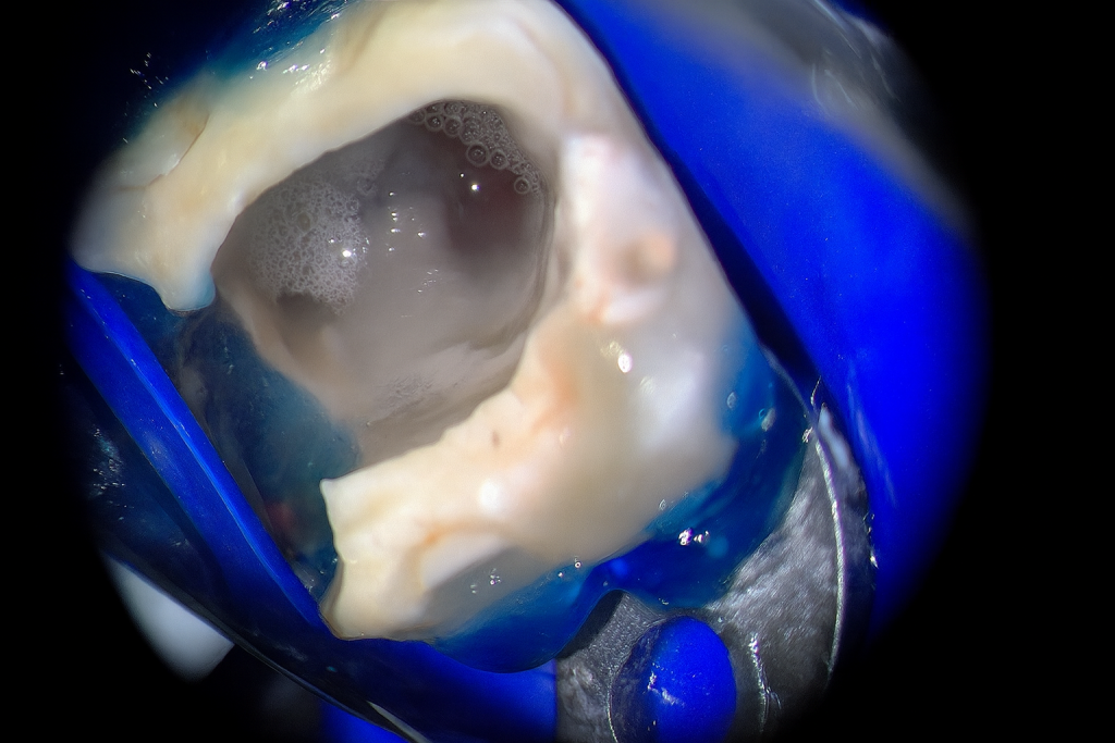
3️⃣ Ledge Bypass and Perforation Management
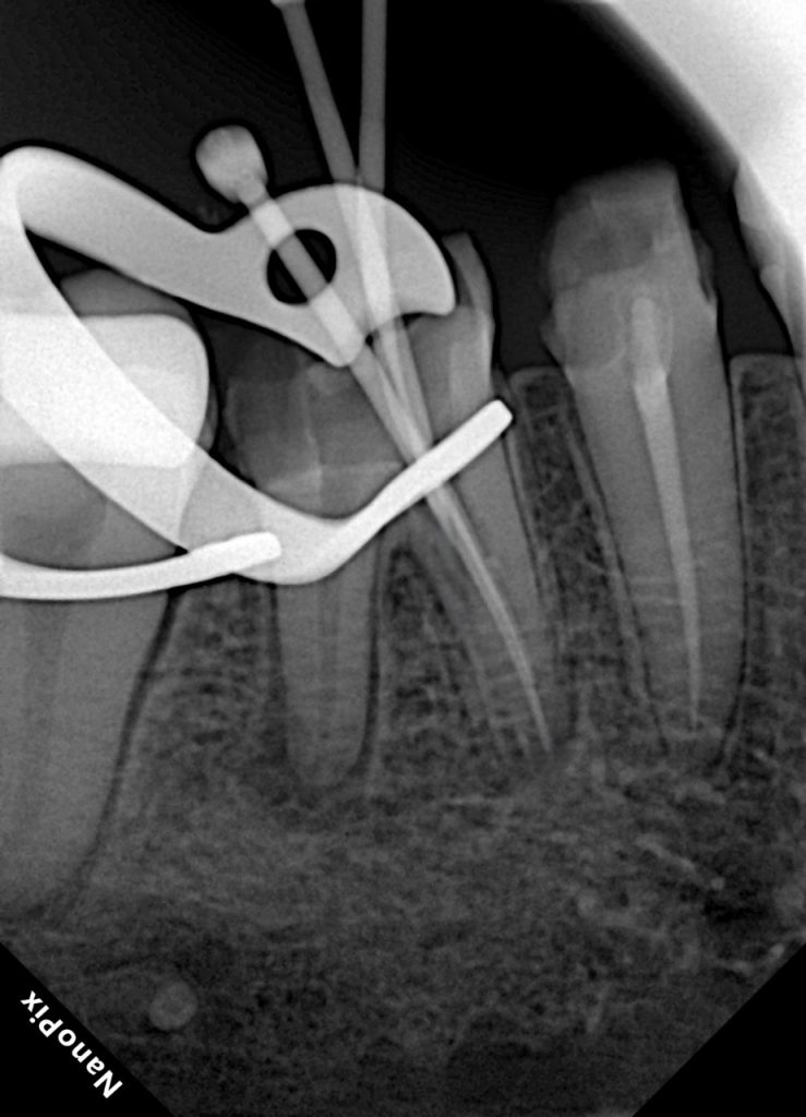
A mechanical ledge was identified in the mesial canal during negotiation. Glide path re-established with #08–10 K-files using watch-winding motion and chelating lubricant (RC-Prep). A small strip perforation was observed and sealed with ProRoot MTA (Fig 3).
4️⃣ Cleaning and Shaping
Root canals shaped with ProTaper Gold F1–F3 under constant irrigation (5.25 % NaOCl + 17 % EDTA) and passive ultrasonic activation. Final rinse with saline and alcohol ensured canal dryness before obturation (Fig 4).
5️⃣ Obturation with Bioceramic Sealer
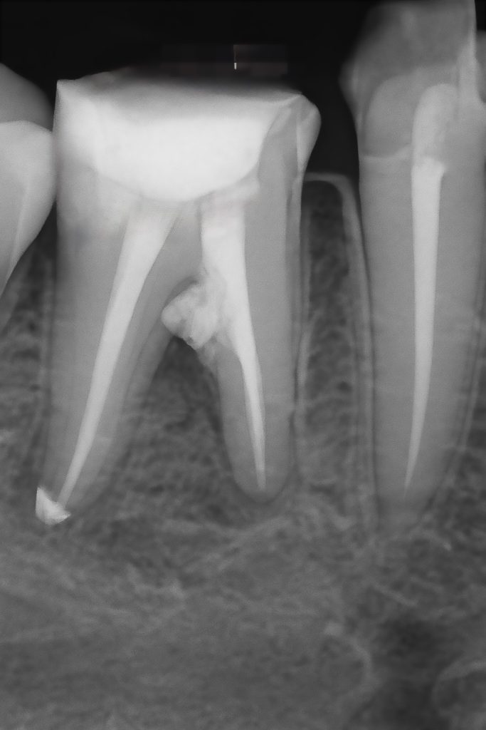
Obturation performed using EndoSequence BC Sealer with single-cone warm vertical compaction. The sealer’s high flow and biomineralization potential provided a tight apical seal and biocompatibility (Fig 5).
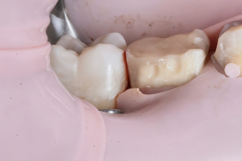
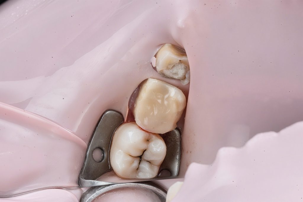
6️⃣ Coronal Reinforcement and Deep Marginal Elevation (DME)
Cervical margin elevated using GC EverX Flow with the matrix-within-matrix technique to re-establish a supragingival margin (Fig 6). Short fibers within the flowable composite acted as an internal stress-absorbing scaffold.
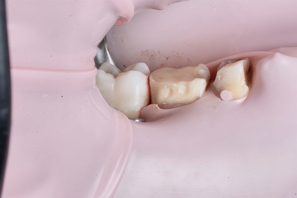
7️⃣ Cuspal Coverage Restoration
Cuspal anatomy reconstructed using Tokuyama Estelite Sigma Quick (A2). Incremental layering with cusp-by-cusp build-up reproduced occlusal morphology and controlled shrinkage. Each increment light-cured for 40 s under microscope visualization (Fig 7).
8️⃣ Finishing & Polishing
Finishing performed with Sof-Lex XT discs, followed by the Lucida Polish System (Style Italiano) to reproduce natural enamel gloss and texture (Fig 8).
9️⃣ Post-operative Evaluation
Radiograph confirmed complete obturation, sealed perforation, and absence of voids (Fig 9). At 1-month recall, the tooth was asymptomatic, functional, and radiographically healing.
Outcome
Through meticulous microscope-guided re-Endo, ultrasonic retrieval, MTA perforation repair, and biomimetic restoration, the tooth was successfully salvaged with full functional and esthetic rehabilitation.
Image Captions
- Fig 1: Pre-op radiograph showing broken file and defective obturation.
- Fig 2: Microscopic access and visualization of fractured instrument.
- Fig 3: Bypassed ledge and MTA placed in perforation area.
- Fig 4: Cleaning and shaping under magnification.
- Fig 5: Obturation with bioceramic sealer.
- Fig 6: DME performed with GC EverX Flow.
- Fig 7: Cuspal build-up with Tokuyama Estelite Sigma Quick.
- Fig 8: Lucida polish for final gloss and texture.
- Fig 9: Post-op radiograph confirming sealing and healing.
Bibliographic References
- Ruddle CJ. Broken Instrument Removal: Strategies for Success. Dent Today 2004; 23(6): 70–73.
- Tay FR, Pashley DH. Monoblocks in Root Canals: A Tangible Goal. J Endod 2007; 33(4): 391–398.
- Mente J et al. Treatment Outcome of Perforations Repaired with Mineral Trioxide Aggregate. J Endod 2010; 36(2): 208–213.
- van Dijken JWV et al. Short Fiber-Reinforced Composites in Posterior Restorations. Dent Mater 2015; 31(5): 545–554.
{"source_type":"hypic","tiktok_developers_3p_anchor_params":"{"capability_key":["edit"],"client_key":"awgvo7gzpeas2ho6","template_id":"","filter_id":[],"capability_extra_v2":{"edit":[{"panel":"hd_quality_picture"},{"panel":"auto_edit_pro"}]}}","data":{"appversion":"7.3.0","product":"retouch","activityName":"","os":"ios","stickerId":"","pictureId":"FF75943A-4ABA-4940-90C3-F0C3B65F3425","playId":"","enter_from":"enter_launch","infoStickerId":"","filterId":"","imageEffectId":""}}
{"tiktok_developers_3p_anchor_params":"{"template_id":"","filter_id":[],"client_key":"awgvo7gzpeas2ho6","capability_key":["edit"],"capability_extra_v2":{"edit":[{"panel":"hd_quality_picture"},{"panel":"auto_edit_pro"}]}}","source_type":"hypic","data":{"stickerId":"","enter_from":"enter_launch","filterId":"","activityName":"","imageEffectId":"","appversion":"7.3.0","infoStickerId":"","os":"ios","pictureId":"2A1320A4-8671-4E1C-ADAD-09506B1ECA19","product":"retouch","playId":""}}
{"source_type":"hypic","tiktok_developers_3p_anchor_params":"{"client_key":"awgvo7gzpeas2ho6","template_id":"","filter_id":[],"capability_extra_v2":{"edit":[{"panel":"hd_quality_picture"},{"panel":"auto_edit_pro"}]},"capability_key":["edit"]}","data":{"appversion":"7.3.0","enter_from":"enter_launch","infoStickerId":"","filterId":"","pictureId":"CD02E653-F630-433A-BB2B-55BE088F30E5","os":"ios","activityName":"","playId":"","product":"retouch","imageEffectId":"","stickerId":""}}
Share on: